Secció de Bioquímica i Biología Molecular, Departament de Bioquímica i Fisiologia, Facultat de Farmàcia i Ciències de l'Alimentació, Universitat de Barcelona, Barcelona, Spain.
Institut de Biomedicina de la Universitat de Barcelona (IBUB), Institut de Recerca Sant Joan de Déu (IRSJD), Barcelona, Spain.
BMC Microbiol. 2019 Jul 17;19(1):166. doi: 10.1186/s12866-019-1534-3.
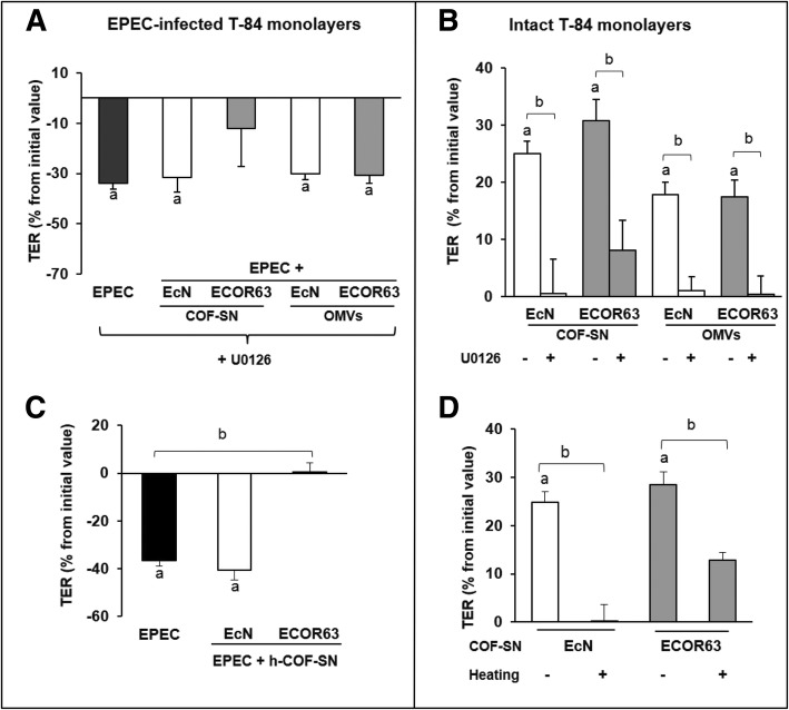
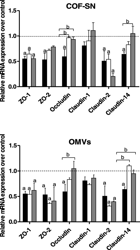
Enteric pathogens have developed mechanisms to disrupt tight junctions and increase gut permeability. Many studies have analysed the ability of live probiotics to protect intestinal epithelial cells against tight junction damage caused by bacterial pathogens. Escherichia coli Nissle 1917 (EcN) is among the probiotics that positively modulates the intestinal epithelial barrier by regulating expression and distribution of tight junction proteins. We previously reported that regulation of ZO-1, claudin-14 and claudin-2 is mediated by EcN secreted factors, either free-released or associated with outer membrane vesicles (OMVs). Factors secreted by commensal ECOR63 elicited comparable effects in intact epithelial T-84 and Caco-2 cell monolayers.
Here we analyse the ability of OMVs and soluble secreted factors to protect epithelial barrier function in polarized T-84 and Caco-2 cells infected with enteropathogenic Escherichia coli (EPEC). Transepithelial electrical resistance, paracellular permeability, mRNA levels and subcellular distribution of tight junction proteins were monitored in the absence or presence of EcN and ECOR63 extracellular fractions. EPEC downregulated expression of ZO-1 ZO-2, occludin and claudin-14 and altered the subcellular localization of ZO-1, occludin and F-actin cytoskeleton. OMVs and soluble factors secreted by EcN and ECOR63 counteracted EPEC-altered transepithelial resistance and paracellular permeability, preserved occludin and claudin-14 mRNA levels, retained ZO-1 and occludin at tight junctions in the cell boundaries and ameliorated F-actin disorganization. Redistribution of ZO-1 was not accompanied by changes at mRNA level.
This study provides new insights on the role of microbiota secreted factors on the modulation of intestinal tight junctions, expanding their barrier-protective effects against pathogen-induced disruption.
肠道病原体已开发出破坏紧密连接并增加肠道通透性的机制。许多研究分析了活益生菌保护肠道上皮细胞免受细菌病原体引起的紧密连接损伤的能力。大肠杆菌 Nissle 1917(EcN)是通过调节紧密连接蛋白的表达和分布来积极调节肠道上皮屏障的益生菌之一。我们之前报道过,EcN 分泌的因子(无论是自由释放的还是与外膜囊泡(OMVs)相关的)调节 ZO-1、claudin-14 和 claudin-2 的调节。共生 ECOR63 分泌的因子在完整的上皮 T-84 和 Caco-2 细胞单层中也产生了类似的效果。
在这里,我们分析了 OMVs 和可溶性分泌因子在感染肠致病性大肠杆菌(EPEC)的极化 T-84 和 Caco-2 细胞中保护上皮屏障功能的能力。在不存在或存在 EcN 和 ECOR63 细胞外部分的情况下,监测跨上皮电阻、旁通透性、紧密连接蛋白的 mRNA 水平和亚细胞分布。EPEC 下调了 ZO-1、ZO-2、occludin 和 claudin-14 的表达,并改变了 ZO-1、occludin 和 F-肌动蛋白细胞骨架的亚细胞定位。EcN 和 ECOR63 分泌的 OMVs 和可溶性因子拮抗了 EPEC 改变的跨上皮电阻和旁通透性,保留了 occludin 和 claudin-14 的 mRNA 水平,保留了紧密连接处的 ZO-1 和 occludin 在细胞边界,并改善了 F-肌动蛋白的紊乱。ZO-1 的重分布没有伴随着 mRNA 水平的变化。
这项研究提供了关于微生物群分泌因子对肠道紧密连接调节作用的新见解,扩展了它们针对病原体诱导的破坏的屏障保护作用。