• 文献检索
  • 文档翻译
  • 深度研究
  • 学术资讯
  • Suppr Zotero 插件Zotero 插件
  • 邀请有礼
  • 套餐&价格
  • 历史记录
应用&插件
Suppr Zotero 插件Zotero 插件浏览器插件Mac 客户端Windows 客户端微信小程序
定价
高级版会员购买积分包购买API积分包
服务
文献检索文档翻译深度研究API 文档MCP 服务
关于我们
关于 Suppr公司介绍联系我们用户协议隐私条款
关注我们

Suppr 超能文献

核心技术专利:CN118964589B侵权必究
粤ICP备2023148730 号-1Suppr @ 2026

文献检索

告别复杂PubMed语法,用中文像聊天一样搜索,搜遍4000万医学文献。AI智能推荐,让科研检索更轻松。

立即免费搜索

文件翻译

保留排版,准确专业,支持PDF/Word/PPT等文件格式,支持 12+语言互译。

免费翻译文档

深度研究

AI帮你快速写综述,25分钟生成高质量综述,智能提取关键信息,辅助科研写作。

立即免费体验

基于不规则年龄图谱的扩张显著度U型网络用于脑白质高信号分割

Dilated Saliency U-Net for White Matter Hyperintensities Segmentation Using Irregularity Age Map.

作者信息

Jeong Yunhee, Rachmadi Muhammad Febrian, Valdés-Hernández Maria Del C, Komura Taku

机构信息

School of Informatics, University of Edinburgh, Edinburgh, United Kingdom.

Centre for Clinical Brain Sciences, University of Edinburgh, Edinburgh, United Kingdom.

出版信息

Front Aging Neurosci. 2019 Jun 27;11:150. doi: 10.3389/fnagi.2019.00150. eCollection 2019.

DOI:10.3389/fnagi.2019.00150
PMID:31316369
原文链接:https://pmc.ncbi.nlm.nih.gov/articles/PMC6610522/
Abstract

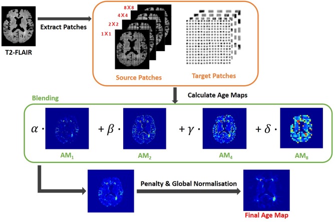
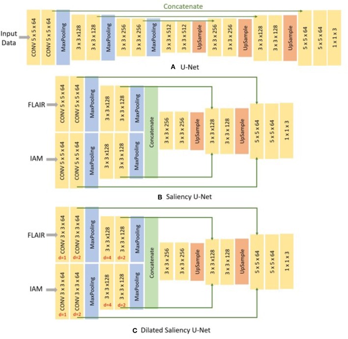
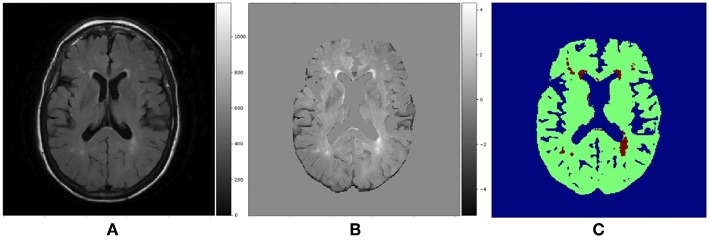
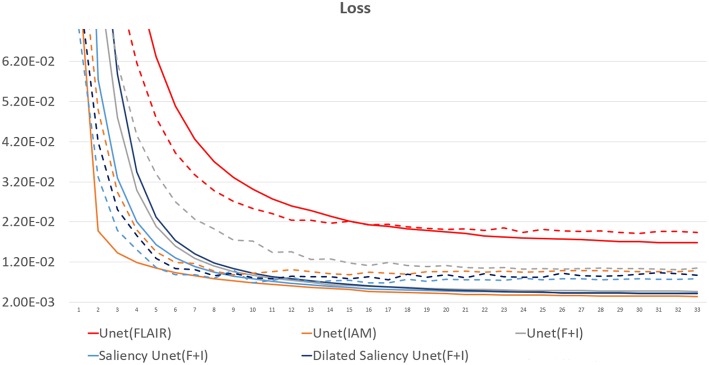
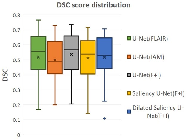
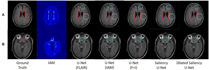
White matter hyperintensities (WMH) appear as regions of abnormally high signal intensity on T2-weighted magnetic resonance image (MRI) sequences. In particular, WMH have been noteworthy in age-related neuroscience for being a crucial biomarker for all types of dementia and brain aging processes. The automatic WMH segmentation is challenging because of their variable intensity range, size and shape. U-Net tackles this problem through the dense prediction and has shown competitive performances not only on WMH segmentation/detection but also on varied image segmentation tasks. However, its network architecture is high complex. In this study, we propose the use of Saliency U-Net and Irregularity map (IAM) to decrease the U-Net architectural complexity without performance loss. We trained Saliency U-Net using both: a T2-FLAIR MRI sequence and its correspondent IAM. Since IAM guides locating image intensity irregularities, in which WMH are possibly included, in the MRI slice, Saliency U-Net performs better than the original U-Net trained only using T2-FLAIR. The best performance was achieved with fewer parameters and shorter training time. Moreover, the application of dilated convolution enhanced Saliency U-Net by recognizing the shape of large WMH more accurately through multi-context learning. This network named Dilated Saliency U-Net improved Dice coefficient score to 0.5588 which was the best score among our experimental models, and recorded a relatively good sensitivity of 0.4747 with the shortest training time and the least number of parameters. In conclusion, based on our experimental results, incorporating IAM through Dilated Saliency U-Net resulted an appropriate approach for WMH segmentation.

摘要

白质高信号(WMH)在T2加权磁共振成像(MRI)序列上表现为异常高信号强度区域。特别是,WMH在与年龄相关的神经科学中一直备受关注,因为它是所有类型痴呆和脑老化过程的关键生物标志物。由于WMH的强度范围、大小和形状各不相同,自动WMH分割具有挑战性。U-Net通过密集预测解决了这个问题,并且不仅在WMH分割/检测方面,而且在各种图像分割任务中都表现出了有竞争力的性能。然而,其网络架构非常复杂。在本研究中,我们提出使用显著性U-Net和不规则性图(IAM)来降低U-Net的架构复杂性,同时不损失性能。我们使用T2-FLAIR MRI序列及其对应的IAM训练显著性U-Net。由于IAM有助于在MRI切片中定位可能包含WMH的图像强度不规则区域,因此显著性U-Net比仅使用T2-FLAIR训练的原始U-Net表现更好。在参数更少和训练时间更短的情况下实现了最佳性能。此外,扩张卷积的应用通过多上下文学习更准确地识别大WMH的形状,增强了显著性U-Net。这个名为扩张显著性U-Net的网络将Dice系数分数提高到了0.5588,这是我们实验模型中的最佳分数,并且在最短的训练时间和最少的参数数量下记录了相对较好的灵敏度0.4747。总之,基于我们的实验结果,通过扩张显著性U-Net结合IAM是一种适用于WMH分割的方法。

https://cdn.ncbi.nlm.nih.gov/pmc/blobs/d89d/6610522/2e7dfa6bb9bb/fnagi-11-00150-g0011.jpg
https://cdn.ncbi.nlm.nih.gov/pmc/blobs/d89d/6610522/5a20c40072e3/fnagi-11-00150-g0001.jpg
https://cdn.ncbi.nlm.nih.gov/pmc/blobs/d89d/6610522/7e26fc102880/fnagi-11-00150-g0002.jpg
https://cdn.ncbi.nlm.nih.gov/pmc/blobs/d89d/6610522/f100bbac4015/fnagi-11-00150-g0003.jpg
https://cdn.ncbi.nlm.nih.gov/pmc/blobs/d89d/6610522/1fed74e44f74/fnagi-11-00150-g0004.jpg
https://cdn.ncbi.nlm.nih.gov/pmc/blobs/d89d/6610522/c94f9daa7824/fnagi-11-00150-g0005.jpg
https://cdn.ncbi.nlm.nih.gov/pmc/blobs/d89d/6610522/0cf5ee8291c9/fnagi-11-00150-g0006.jpg
https://cdn.ncbi.nlm.nih.gov/pmc/blobs/d89d/6610522/3123193ad08f/fnagi-11-00150-g0007.jpg
https://cdn.ncbi.nlm.nih.gov/pmc/blobs/d89d/6610522/add05714898a/fnagi-11-00150-g0008.jpg
https://cdn.ncbi.nlm.nih.gov/pmc/blobs/d89d/6610522/10ceaae87279/fnagi-11-00150-g0009.jpg
https://cdn.ncbi.nlm.nih.gov/pmc/blobs/d89d/6610522/c524cce6009a/fnagi-11-00150-g0010.jpg
https://cdn.ncbi.nlm.nih.gov/pmc/blobs/d89d/6610522/2e7dfa6bb9bb/fnagi-11-00150-g0011.jpg

相似文献

1
Dilated Saliency U-Net for White Matter Hyperintensities Segmentation Using Irregularity Age Map.基于不规则年龄图谱的扩张显著度U型网络用于脑白质高信号分割
Front Aging Neurosci. 2019 Jun 27;11:150. doi: 10.3389/fnagi.2019.00150. eCollection 2019.
2
Automatic segmentation of white matter hyperintensities: validation and comparison with state-of-the-art methods on both Multiple Sclerosis and elderly subjects.自动分割脑白质高信号:在多发性硬化症和老年患者中验证和比较最先进的方法。
Neuroimage Clin. 2022;33:102940. doi: 10.1016/j.nicl.2022.102940. Epub 2022 Jan 10.
3
Limited One-time Sampling Irregularity Map (LOTS-IM) for Automatic Unsupervised Assessment of White Matter Hyperintensities and Multiple Sclerosis Lesions in Structural Brain Magnetic Resonance Images.用于结构磁共振成像中白质高信号和多发性硬化病变自动无监督评估的有限一次性采样不规则图(LOTS-IM)。
Comput Med Imaging Graph. 2020 Jan;79:101685. doi: 10.1016/j.compmedimag.2019.101685. Epub 2019 Nov 27.
4
Automatic segmentation of white matter hyperintensities in T2-FLAIR with AQUA: A comparative validation study against conventional methods.使用AQUA对T2-FLAIR序列中的脑白质高信号进行自动分割:与传统方法的比较验证研究
Brain Res Bull. 2023 Dec;205:110825. doi: 10.1016/j.brainresbull.2023.110825. Epub 2023 Nov 22.
5
Multi-Disease Segmentation of Gliomas and White Matter Hyperintensities in the BraTS Data Using a 3D Convolutional Neural Network.使用3D卷积神经网络对BraTS数据中的胶质瘤和脑白质高信号进行多疾病分割
Front Comput Neurosci. 2019 Dec 20;13:84. doi: 10.3389/fncom.2019.00084. eCollection 2019.
6
White matter hyperintensities segmentation using the ensemble U-Net with multi-scale highlighting foregrounds.使用多尺度高亮前景的集成 U-Net 进行脑白质高信号分割。
Neuroimage. 2021 Aug 15;237:118140. doi: 10.1016/j.neuroimage.2021.118140. Epub 2021 May 3.
7
Specialized gray matter segmentation via a generative adversarial network: application on brain white matter hyperintensities classification.通过生成对抗网络进行的专门灰质分割:在脑白质高信号分类中的应用。
Front Neurosci. 2024 Sep 30;18:1416174. doi: 10.3389/fnins.2024.1416174. eCollection 2024.
8
Segmentation and differentiation of periventricular and deep white matter hyperintensities in 2D T2-FLAIR MRI based on a cascade U-net.基于级联U型网络的二维T2液体衰减反转恢复序列磁共振成像中脑室周围和深部白质高信号的分割与鉴别
Front Neurol. 2022 Nov 17;13:1021477. doi: 10.3389/fneur.2022.1021477. eCollection 2022.
9
An anatomical knowledge-based MRI deep learning pipeline for white matter hyperintensity quantification associated with cognitive impairment.基于解剖学知识的 MRI 深度学习流水线用于量化与认知障碍相关的脑白质高信号。
Comput Med Imaging Graph. 2021 Apr;89:101873. doi: 10.1016/j.compmedimag.2021.101873. Epub 2021 Feb 3.
10
Intra-Scanner and Inter-Scanner Reproducibility of Automatic White Matter Hyperintensities Quantification.自动脑白质高信号定量的扫描仪内及扫描仪间的可重复性
Front Neurosci. 2019 Jul 10;13:679. doi: 10.3389/fnins.2019.00679. eCollection 2019.

引用本文的文献

1
White matter hyperintensities segmentation using an ensemble of neural networks.基于神经网络集成的脑白质高信号分割。
Hum Brain Mapp. 2022 Feb 15;43(3):929-939. doi: 10.1002/hbm.25695. Epub 2021 Oct 27.
2
Using Artificial Intelligence for Automatic Segmentation of CT Lung Images in Acute Respiratory Distress Syndrome.利用人工智能对急性呼吸窘迫综合征的CT肺图像进行自动分割
Front Physiol. 2021 Sep 14;12:676118. doi: 10.3389/fphys.2021.676118. eCollection 2021.
3
Automatic spatial estimation of white matter hyperintensities evolution in brain MRI using disease evolution predictor deep neural networks.

本文引用的文献

1
Limited One-time Sampling Irregularity Map (LOTS-IM) for Automatic Unsupervised Assessment of White Matter Hyperintensities and Multiple Sclerosis Lesions in Structural Brain Magnetic Resonance Images.用于结构磁共振成像中白质高信号和多发性硬化病变自动无监督评估的有限一次性采样不规则图(LOTS-IM)。
Comput Med Imaging Graph. 2020 Jan;79:101685. doi: 10.1016/j.compmedimag.2019.101685. Epub 2019 Nov 27.
2
Brain lesion segmentation through image synthesis and outlier detection.通过图像合成和异常值检测进行脑损伤分割。
Neuroimage Clin. 2017 Sep 8;16:643-658. doi: 10.1016/j.nicl.2017.09.003. eCollection 2017.
3
使用疾病进展预测深度神经网络对脑部磁共振成像中白质高信号演变进行自动空间估计。
Med Image Anal. 2020 Jul;63:101712. doi: 10.1016/j.media.2020.101712. Epub 2020 Apr 26.
4
Deep Learning and Neurology: A Systematic Review.深度学习与神经病学:一项系统综述。
Neurol Ther. 2019 Dec;8(2):351-365. doi: 10.1007/s40120-019-00153-8. Epub 2019 Aug 21.
White matter hyperintensity and stroke lesion segmentation and differentiation using convolutional neural networks.
基于卷积神经网络的脑白质高信号和卒中病灶分割与区分。
Neuroimage Clin. 2017 Dec 20;17:918-934. doi: 10.1016/j.nicl.2017.12.022. eCollection 2018.
4
Fully Convolutional Networks for Semantic Segmentation.全卷积网络用于语义分割。
IEEE Trans Pattern Anal Mach Intell. 2017 Apr;39(4):640-651. doi: 10.1109/TPAMI.2016.2572683. Epub 2016 May 24.
5
Multiple-Instance Learning for Anomaly Detection in Digital Mammography.基于多示例学习的数字乳腺图像异常检测
IEEE Trans Med Imaging. 2016 Jul;35(7):1604-14. doi: 10.1109/TMI.2016.2521442. Epub 2016 Jan 25.
6
Rationale, design and methodology of the image analysis protocol for studies of patients with cerebral small vessel disease and mild stroke.用于脑小血管病和轻度中风患者研究的图像分析方案的原理、设计和方法。
Brain Behav. 2015 Nov 26;5(12):e00415. doi: 10.1002/brb3.415. eCollection 2015 Dec.
7
Optimized brain extraction for pathological brains (optiBET).针对病理性大脑的优化脑提取(optiBET)。
PLoS One. 2014 Dec 16;9(12):e115551. doi: 10.1371/journal.pone.0115551. eCollection 2014.
8
Volume of white matter hyperintensities in healthy adults: contribution of age, vascular risk factors, and inflammation-related genetic variants.健康成年人脑白质高信号体积:年龄、血管危险因素及炎症相关基因变异的作用
Biochim Biophys Acta. 2012 Mar;1822(3):361-9. doi: 10.1016/j.bbadis.2011.08.007. Epub 2011 Aug 25.
9
Evaluating intensity normalization on MRIs of human brain with multiple sclerosis.评估多发性硬化症患者脑部 MRI 的强度归一化。
Med Image Anal. 2011 Apr;15(2):267-82. doi: 10.1016/j.media.2010.12.003. Epub 2010 Dec 25.
10
Neuropathological basis of magnetic resonance images in aging and dementia.衰老和痴呆中磁共振成像的神经病理学基础。
Ann Neurol. 2008 Jan;63(1):72-80. doi: 10.1002/ana.21296.