Guo Chunjie, Niu Kai, Luo Yishan, Shi Lin, Wang Zhuo, Zhao Meng, Wang Defeng, Zhu Wan'an, Zhang Huimao, Sun Li
Department of Radiology, The First Hospital of Jilin University, Changchun, China.
Department of Otorhinolaryngology Head and Neck Surgery, The First Hospital of Jilin University, Changchun, China.
Front Neurosci. 2019 Jul 10;13:679. doi: 10.3389/fnins.2019.00679. eCollection 2019.
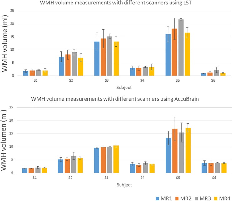
To evaluate white matter hyperintensities (WMH) quantification reproducibility from multiple aspects of view and examine the effects of scan-rescan procedure, types of scanner, imaging protocols, scanner software upgrade, and automatic segmentation tools on WMH quantification results using magnetic resonance imaging (MRI). Six post-stroke subjects (4 males; mean age = 62.8, range = 58-72 years) were scanned and rescanned with both 3D T1-weighted, 2D and 3D T2-weighted fluid-attenuated inversion recovery (T2-FLAIR) MRI across four different MRI scanners within 12 h. Two automated WMH segmentation and quantification tools were used to measure WMH volume based on each MR scan. Robustness was assessed using the coefficient of variation (CV), Dice similarity coefficient (DSC), and intra-class correlation (ICC). Experimental results show that the best reproducibility was achieved by using 3D T2-FLAIR MRI under intra-scanner setting with CV ranging from 2.69 to 2.97%, while the largest variability resulted from comparing WMH volumes measured based on 2D T2-FLAIR MRI with those of 3D T2-FLAIR MRI, with CV values in the range of 15.62%-29.33%. The WMH quantification variability based on 2D MRIs is larger than 3D MRIs due to their large slice thickness. The DSC of WMH segmentation labels between intra-scanner MRIs ranges from 0.63 to 0.77, while that for inter-scanner MRIs is in the range of 0.63-0.65. In addition to image acquisition, the choice of automatic WMH segmentation tool also has a large impact on WMH quantification. WMH reproducibility is one of the primary issues to be considered in multicenter and longitudinal studies. The study provides solid guidance in assisting multicenter and longitudinal study design to achieve meaningful results with enough power.
从多个视角评估脑白质高信号(WMH)定量的可重复性,并研究扫描 - 重扫程序、扫描仪类型、成像协议、扫描仪软件升级以及自动分割工具对使用磁共振成像(MRI)进行WMH定量结果的影响。对6名中风后受试者(4名男性;平均年龄 = 62.8岁,范围 = 58 - 72岁)在12小时内使用四台不同的MRI扫描仪进行3D T1加权、2D和3D T2加权液体衰减反转恢复(T2 - FLAIR)MRI扫描和重扫。使用两种自动WMH分割和定量工具基于每次MR扫描测量WMH体积。使用变异系数(CV)、骰子相似系数(DSC)和组内相关性(ICC)评估稳健性。实验结果表明,在扫描仪内设置下使用3D T2 - FLAIR MRI可实现最佳可重复性,CV范围为2.69%至2.97%,而基于2D T2 - FLAIR MRI测量的WMH体积与3D T2 - FLAIR MRI测量的WMH体积相比,变异最大,CV值在15.62% - 29.33%范围内。由于2D MRI的切片厚度较大,基于2D MRI的WMH定量变异性大于3D MRI。扫描仪内MRI之间WMH分割标签的DSC范围为0.63至0.77,而扫描仪间MRI的DSC范围为0.63 - 0.65。除了图像采集外,自动WMH分割工具的选择对WMH定量也有很大影响。WMH可重复性是多中心和纵向研究中要考虑的主要问题之一。该研究为协助多中心和纵向研究设计以获得具有足够效力的有意义结果提供了坚实的指导。