Lutkenhoff Evan S, Rosenberg Matthew, Chiang Jeffrey, Zhang Kunyu, Pickard John D, Owen Adrian M, Monti Martin M
Department of Psychology, University of California Los Angeles, Los Angeles, California, United States of America.
Division of Neurosurgery, University of Cambridge, Addenbrooke's Hospital, Cambridge, United Kingdom.
PLoS One. 2014 Dec 16;9(12):e115551. doi: 10.1371/journal.pone.0115551. eCollection 2014.
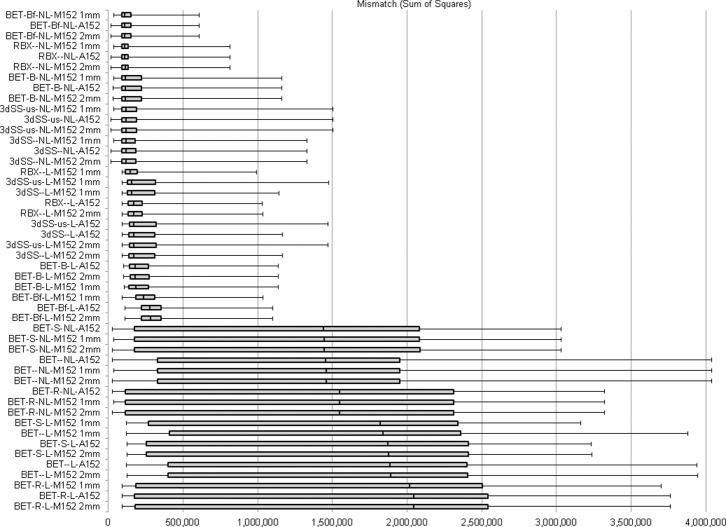
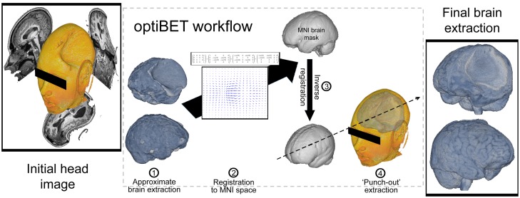
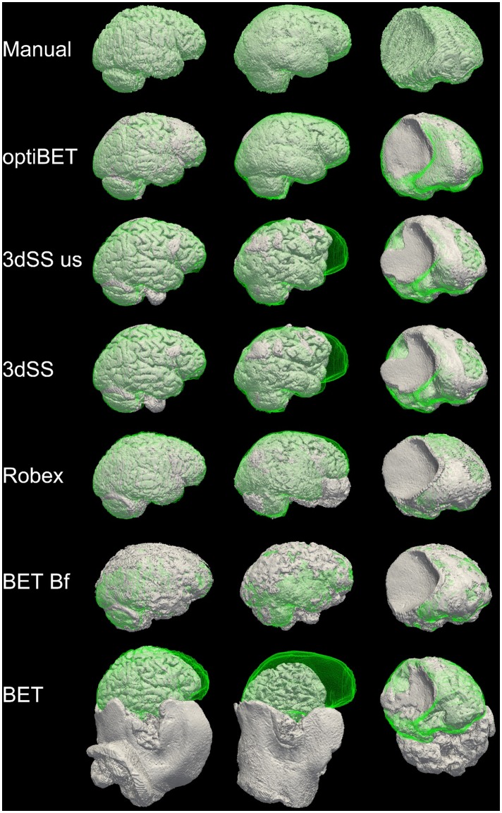
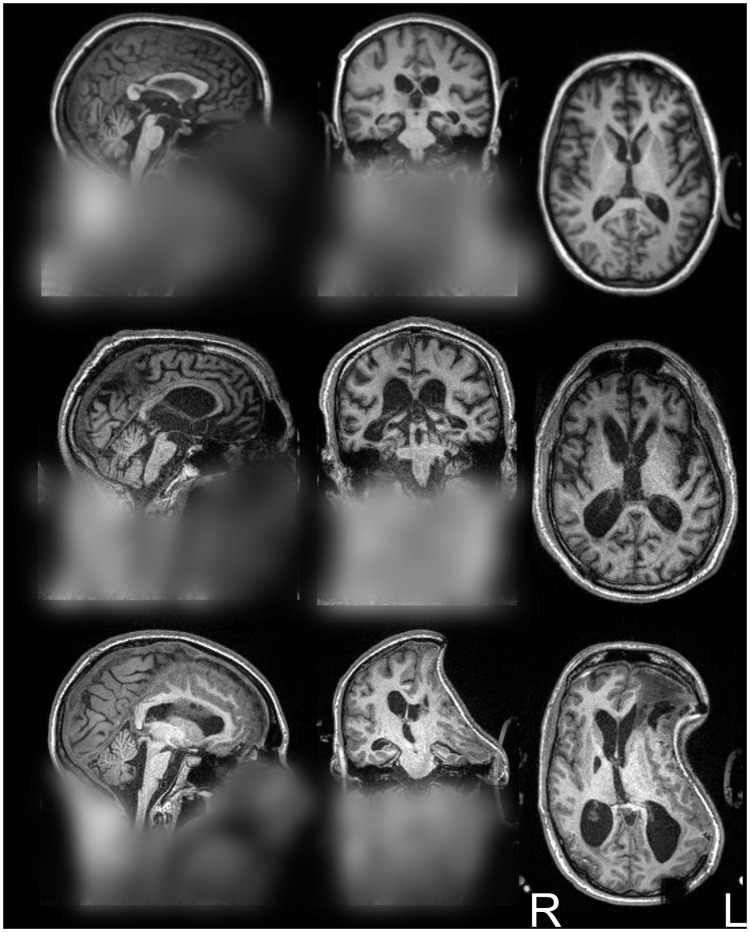
The study of structural and functional magnetic resonance imaging data has greatly benefitted from the development of sophisticated and efficient algorithms aimed at automating and optimizing the analysis of brain data. We address, in the context of the segmentation of brain from non-brain tissue (i.e., brain extraction, also known as skull-stripping), the tension between the increased theoretical and clinical interest in patient data, and the difficulty of conventional algorithms to function optimally in the presence of gross brain pathology. Indeed, because of the reliance of many algorithms on priors derived from healthy volunteers, images with gross pathology can severely affect their ability to correctly trace the boundaries between brain and non-brain tissue, potentially biasing subsequent analysis. We describe and make available an optimized brain extraction script for the pathological brain (optiBET) robust to the presence of pathology. Rather than attempting to trace the boundary between tissues, optiBET performs brain extraction by (i) calculating an initial approximate brain extraction; (ii) employing linear and non-linear registration to project the approximate extraction into the MNI template space; (iii) back-projecting a standard brain-only mask from template space to the subject's original space; and (iv) employing the back-projected brain-only mask to mask-out non-brain tissue. The script results in up to 94% improvement of the quality of extractions over those obtained with conventional software across a large set of severely pathological brains. Since optiBET makes use of freely available algorithms included in FSL, it should be readily employable by anyone having access to such tools.
复杂高效算法的发展极大地推动了对结构和功能磁共振成像数据的研究,这些算法旨在实现脑数据分析的自动化并进行优化。在从非脑组织中分割出脑(即脑提取,也称为去颅骨)的背景下,我们探讨了对患者数据在理论和临床方面日益增长的兴趣与传统算法在存在严重脑病变时难以实现最佳功能之间的矛盾。实际上,由于许多算法依赖于从健康志愿者得出的先验信息,存在严重病变的图像会严重影响它们正确描绘脑与非脑组织之间边界的能力,这可能会使后续分析产生偏差。我们描述并提供了一种针对病变脑的优化脑提取脚本(optiBET),它对病变具有鲁棒性。optiBET不是试图追踪组织之间的边界,而是通过以下方式进行脑提取:(i)计算初始的近似脑提取;(ii)采用线性和非线性配准将近似提取投影到MNI模板空间;(iii)将仅包含脑的标准掩码从模板空间反投影到受试者的原始空间;(iv)使用反投影的仅包含脑的掩码来掩盖非脑组织。在一大组严重病变的脑图像上,该脚本生成的提取质量比使用传统软件得到的结果提高了多达94%。由于optiBET利用了FSL中包含的免费算法,任何能够使用这些工具的人都应该能够轻松使用它。