• 文献检索
  • 文档翻译
  • 深度研究
  • 学术资讯
  • Suppr Zotero 插件Zotero 插件
  • 邀请有礼
  • 套餐&价格
  • 历史记录
应用&插件
Suppr Zotero 插件Zotero 插件浏览器插件Mac 客户端Windows 客户端微信小程序
定价
高级版会员购买积分包购买API积分包
服务
文献检索文档翻译深度研究API 文档MCP 服务
关于我们
关于 Suppr公司介绍联系我们用户协议隐私条款
关注我们

Suppr 超能文献

核心技术专利:CN118964589B侵权必究
粤ICP备2023148730 号-1Suppr @ 2026

文献检索

告别复杂PubMed语法,用中文像聊天一样搜索,搜遍4000万医学文献。AI智能推荐,让科研检索更轻松。

立即免费搜索

文件翻译

保留排版,准确专业,支持PDF/Word/PPT等文件格式,支持 12+语言互译。

免费翻译文档

深度研究

AI帮你快速写综述,25分钟生成高质量综述,智能提取关键信息,辅助科研写作。

立即免费体验

应用超微血管成像技术评估小儿肠系膜淋巴结炎患儿的肠系膜淋巴结。

Evaluation of Mesenteric Lymph Nodes in a Pediatric Population with Mesenteric Lymphadenitis Using Superb Microvascular Imaging.

机构信息

Department of Pediatrics, Pudong New Area Peoples' Hospital Affiliated to Shanghai University of Medicine and Health Sciences, Shanghai, China (mainland).

Department of Gynecology and Obstetrics, Pudong New Area Peoples' Hospital Affiliated to Shanghai University of Medicine and Health Sciences, Shanghai, China (mainland).

出版信息

Med Sci Monit. 2019 Jul 18;25:5336-5342. doi: 10.12659/MSM.914903.

DOI:10.12659/MSM.914903
PMID:31318849
原文链接:https://pmc.ncbi.nlm.nih.gov/articles/PMC6659467/
Abstract

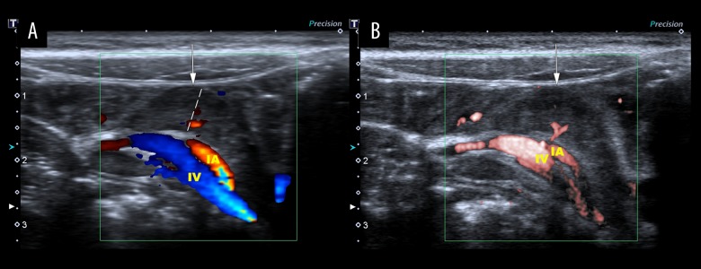
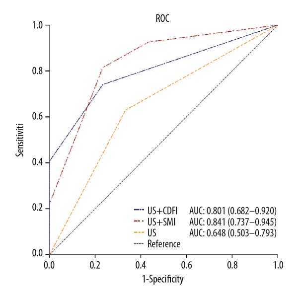
BACKGROUND This study aimed to evaluate superb microvascular imaging (SMI) as an adjunctive imaging method to evaluate mesenteric lymph nodes in children with mesenteric lymphadenitis compared with healthy children. MATERIAL AND METHODS A retrospective study compared children with mesenteric lymphadenitis (n=27) and healthy children (n=30). Lymph node size was determined using grayscale ultrasonography and parameters of lymph node vascularity were compared using color Doppler flow imaging (CDFI) and SMI. The diagnostic performance of ultrasound (US), US combined with SMI, and US combined with CDFI were compared. RESULTS Lymph nodes from children with mesenteric lymphadenitis (n=77) and normal lymph nodes (n=84) were evaluated by SMI, which showed that the least diameter of lymph nodes in cases of mesenteric lymphadenitis was 0.58±0.15 mm and of normal mesenteric lymph nodes was 0.47±0.08 mm (p<0.001). SMI identified 92.6% of abnormal mesenteric lymph nodes while CDFI detected 85.2%. US combined with SMI had the highest sensitivity (81.5%), and specificity (78.9%) compared with US alone (sensitivity, 63.0%; specificity, 64.9%), and compared with US combined with CDFI (sensitivity, 74.1%; specificity, 75.4%). US combined with SMI and US combined with CDFI achieved the same specificity (76.7%), which was higher than that of US alone (66.7%). CONCLUSIONS SMI was superior to color Doppler flow imaging in evaluating the microvasculature in lymphadenopathy in mesenteric lymphadenitis. SMI may be used as an adjunct to grayscale ultrasonography to assist in identifying mesenteric lymphadenopathy in pediatric patients.

摘要

背景

本研究旨在评估超级微血管成像(SMI)作为一种辅助成像方法,用于评估肠系膜淋巴结炎患儿与健康儿童的肠系膜淋巴结,与健康儿童相比。

材料与方法

回顾性研究比较了肠系膜淋巴结炎患儿(n=27)和健康儿童(n=30)。使用灰阶超声测量淋巴结大小,并比较彩色多普勒血流成像(CDFI)和 SMI 评估淋巴结血管参数。比较超声(US)、US 联合 SMI 和 US 联合 CDFI 的诊断性能。

结果

肠系膜淋巴结炎患儿(n=77)和正常肠系膜淋巴结(n=84)的淋巴结进行了 SMI 评估,结果显示肠系膜淋巴结炎患儿的淋巴结最小直径为 0.58±0.15mm,正常肠系膜淋巴结为 0.47±0.08mm(p<0.001)。SMI 可识别 92.6%的异常肠系膜淋巴结,而 CDFI 可识别 85.2%。与单独使用 US(敏感性,63.0%;特异性,64.9%)相比,US 联合 SMI 的敏感性(81.5%)和特异性(78.9%)最高,与 US 联合 CDFI(敏感性,74.1%;特异性,75.4%)相比。US 联合 SMI 和 US 联合 CDFI 的特异性相同(76.7%),高于单独使用 US(66.7%)。

结论

SMI 在评估肠系膜淋巴结炎淋巴结病变的微血管方面优于彩色多普勒血流成像。SMI 可作为灰阶超声的辅助手段,用于辅助识别儿科患者的肠系膜淋巴结病变。

https://cdn.ncbi.nlm.nih.gov/pmc/blobs/b85f/6659467/8bec10472d52/medscimonit-25-5336-g003.jpg
https://cdn.ncbi.nlm.nih.gov/pmc/blobs/b85f/6659467/688c8a5923f6/medscimonit-25-5336-g001.jpg
https://cdn.ncbi.nlm.nih.gov/pmc/blobs/b85f/6659467/904bb1f931c2/medscimonit-25-5336-g002.jpg
https://cdn.ncbi.nlm.nih.gov/pmc/blobs/b85f/6659467/8bec10472d52/medscimonit-25-5336-g003.jpg
https://cdn.ncbi.nlm.nih.gov/pmc/blobs/b85f/6659467/688c8a5923f6/medscimonit-25-5336-g001.jpg
https://cdn.ncbi.nlm.nih.gov/pmc/blobs/b85f/6659467/904bb1f931c2/medscimonit-25-5336-g002.jpg
https://cdn.ncbi.nlm.nih.gov/pmc/blobs/b85f/6659467/8bec10472d52/medscimonit-25-5336-g003.jpg

相似文献

1
Evaluation of Mesenteric Lymph Nodes in a Pediatric Population with Mesenteric Lymphadenitis Using Superb Microvascular Imaging.应用超微血管成像技术评估小儿肠系膜淋巴结炎患儿的肠系膜淋巴结。
Med Sci Monit. 2019 Jul 18;25:5336-5342. doi: 10.12659/MSM.914903.
2
A Prospective Study to Compare Superb Microvascular Imaging with Grayscale Ultrasound and Color Doppler Flow Imaging of Vascular Distribution and Morphology in Thyroid Nodules.一项对比甲状腺结节血管分布和形态的灰阶超声及彩色多普勒血流显像与超级微血管成像的前瞻性研究。
Med Sci Monit. 2018 Dec 19;24:9223-9231. doi: 10.12659/MSM.911695.
3
Diagnostic performances of superb microvascular imaging, shear wave elastography and shape index in pediatric lymph nodes categorization: a comparative study.超微血管成像、剪切波弹性成像及形状指数在儿童淋巴结分类中的诊断性能:一项对比研究
Br J Radiol. 2018 Jul;91(1087):20180129. doi: 10.1259/bjr.20180129. Epub 2018 May 11.
4
Diagnostic Performance of Superb Microvascular Imaging (SMI) Combined with Shear-Wave Elastography in Evaluating Breast Lesions.超声微血管成像(SMI)联合剪切波弹性成像对乳腺病变的诊断性能评估。
Med Sci Monit. 2018 Aug 26;24:5935-5942. doi: 10.12659/MSM.910399.
5
Use superb microvascular imaging to diagnose and predict metastatic cervical lymph nodes in patients with papillary thyroid carcinoma.运用高超的微血管成像技术诊断和预测甲状腺乳头状癌患者转移性颈淋巴结。
J Cancer Res Clin Oncol. 2024 May 21;150(5):268. doi: 10.1007/s00432-024-05770-x.
6
Differentiation Between Malignant and Benign Lymph Nodes: Role of Superb Microvascular Imaging in the Evaluation of Cervical Lymph Nodes.良恶性淋巴结的鉴别:超微血管成像在评估颈部淋巴结中的作用。
J Ultrasound Med. 2019 Nov;38(11):3025-3036. doi: 10.1002/jum.15010. Epub 2019 Apr 3.
7
[Application value of superb microvascular imaging for diagnosis of different size renal solid tumors].[超微血管成像在不同大小肾实性肿瘤诊断中的应用价值]
Zhonghua Zhong Liu Za Zhi. 2021 Nov 23;43(11):1215-1221. doi: 10.3760/cma.j.cn112152-20191220-00832.
8
Clinical applications of superb microvascular imaging and virtual touch imaging quantification in pediatric mesenteric lymphadenitis diagnosis: A promising pathway to enhanced precision.超微血管成像和虚拟触诊成像定量技术在小儿肠系膜淋巴结炎诊断中的临床应用:提高诊断精准度的一条有前景的途径
Clin Hemorheol Microcirc. 2024;88(4):443-454. doi: 10.3233/CH-242305.
9
The comparative study of color doppler flow imaging, superb microvascular imaging, contrast-enhanced ultrasound micro flow imaging in blood flow analysis of solid renal mass.彩色多普勒血流成像、超微血流成像、超声造影微血流成像在实性肾肿块血流分析中的对比研究。
Cancer Imaging. 2022 May 3;22(1):21. doi: 10.1186/s40644-022-00458-2.
10
Comparison of Doppler Imaging and Microvascular Imaging in Cervical Lymph Node Blood Flow Analysis.多普勒成像与微血管成像在颈淋巴结血流分析中的比较。
Curr Med Imaging. 2024;20(1):e15734056306197. doi: 10.2174/0115734056306197240404035009.

引用本文的文献

1
B-flow imaging in abdominal ultrasound: a pictorial essay.腹部超声中的B-flow成像:图文综述
J Ultrasound. 2025 Aug 21. doi: 10.1007/s40477-025-01068-x.
2
Ultrasound innovations in abdominal radiology: techniques and clinical applications in pediatric imaging.腹部放射学中的超声创新:儿科影像中的技术与临床应用
Abdom Radiol (NY). 2025 Apr;50(4):1744-1762. doi: 10.1007/s00261-024-04616-x. Epub 2024 Oct 16.
3
Clinical applications of superb microvascular imaging and virtual touch imaging quantification in pediatric mesenteric lymphadenitis diagnosis: A promising pathway to enhanced precision.

本文引用的文献

1
A Prospective Study to Compare Superb Microvascular Imaging with Grayscale Ultrasound and Color Doppler Flow Imaging of Vascular Distribution and Morphology in Thyroid Nodules.一项对比甲状腺结节血管分布和形态的灰阶超声及彩色多普勒血流显像与超级微血管成像的前瞻性研究。
Med Sci Monit. 2018 Dec 19;24:9223-9231. doi: 10.12659/MSM.911695.
2
Evaluating the Efficacy of Atorvastatin on Patients with Carotid Plaque by an Innovative Ultrasonography.采用创新超声检查法评估阿托伐他汀对颈动脉斑块患者的疗效。
J Stroke Cerebrovasc Dis. 2019 Mar;28(3):830-837. doi: 10.1016/j.jstrokecerebrovasdis.2018.11.027. Epub 2018 Dec 15.
3
超微血管成像和虚拟触诊成像定量技术在小儿肠系膜淋巴结炎诊断中的临床应用:提高诊断精准度的一条有前景的途径
Clin Hemorheol Microcirc. 2024;88(4):443-454. doi: 10.3233/CH-242305.
Diagnostic performances of superb microvascular imaging, shear wave elastography and shape index in pediatric lymph nodes categorization: a comparative study.
超微血管成像、剪切波弹性成像及形状指数在儿童淋巴结分类中的诊断性能:一项对比研究
Br J Radiol. 2018 Jul;91(1087):20180129. doi: 10.1259/bjr.20180129. Epub 2018 May 11.
4
Superb Microvascular Imaging: Added Value and Novel Applications.超微血管成像:附加价值与新应用
J Clin Imaging Sci. 2017 Dec 28;7:45. doi: 10.4103/jcis.JCIS_79_17. eCollection 2017.
5
US Emergency Department Trends in Imaging for Pediatric Nontraumatic Abdominal Pain.美国急诊科儿科非创伤性腹痛影像学检查的趋势。
Pediatrics. 2017 Oct;140(4). doi: 10.1542/peds.2017-0615. Epub 2017 Sep 15.
6
Acute Nonspecific Mesenteric Lymphadenitis: More Than "No Need for Surgery".急性非特异性肠系膜淋巴结炎:不止是“无需手术”
Biomed Res Int. 2017;2017:9784565. doi: 10.1155/2017/9784565. Epub 2017 Feb 2.
7
A New Era in Diagnostic Ultrasound, Superb Microvascular Imaging: Preliminary Results in Pediatric Hepato-Gastrointestinal Disorders.诊断超声的新时代,卓越的微血管成像:小儿肝胃肠疾病的初步结果
Eur J Pediatr Surg. 2017 Feb;27(1):20-25. doi: 10.1055/s-0036-1593381. Epub 2016 Oct 3.
8
Color Doppler Imaging of the Appendix: Criteria to Improve Specificity for Appendicitis in the Borderline-Size Appendix.阑尾的彩色多普勒成像:提高临界大小阑尾阑尾炎诊断特异性的标准。
J Ultrasound Med. 2016 Oct;35(10):2129-38. doi: 10.7863/ultra.15.11064. Epub 2016 Aug 25.
9
Comparison of the superb microvascular imaging technique and the color Doppler techniques for evaluating children's testicular blood flow.用于评估儿童睾丸血流的超微血管成像技术与彩色多普勒技术的比较。
Eur Rev Med Pharmacol Sci. 2016 May;20(10):1947-53.
10
Comparison of Superb Micro-Vascular Ultrasound Imaging (SMI) and Contrast-Enhanced Ultrasound (CEUS) for Detection of Endoleaks After Endovascular Aneurysm Repair (EVAR).用于检测血管内动脉瘤修复术(EVAR)后内漏的超微血管超声成像(SMI)与对比增强超声(CEUS)的比较
Am J Case Rep. 2016 Jan 25;17:43-6. doi: 10.12659/ajcr.895415.