• 文献检索
  • 文档翻译
  • 深度研究
  • 学术资讯
  • Suppr Zotero 插件Zotero 插件
  • 邀请有礼
  • 套餐&价格
  • 历史记录
应用&插件
Suppr Zotero 插件Zotero 插件浏览器插件Mac 客户端Windows 客户端微信小程序
定价
高级版会员购买积分包购买API积分包
服务
文献检索文档翻译深度研究API 文档MCP 服务
关于我们
关于 Suppr公司介绍联系我们用户协议隐私条款
关注我们

Suppr 超能文献

核心技术专利:CN118964589B侵权必究
粤ICP备2023148730 号-1Suppr @ 2026

文献检索

告别复杂PubMed语法,用中文像聊天一样搜索,搜遍4000万医学文献。AI智能推荐,让科研检索更轻松。

立即免费搜索

文件翻译

保留排版,准确专业,支持PDF/Word/PPT等文件格式,支持 12+语言互译。

免费翻译文档

深度研究

AI帮你快速写综述,25分钟生成高质量综述,智能提取关键信息,辅助科研写作。

立即免费体验

超声微血管成像(SMI)联合剪切波弹性成像对乳腺病变的诊断性能评估。

Diagnostic Performance of Superb Microvascular Imaging (SMI) Combined with Shear-Wave Elastography in Evaluating Breast Lesions.

机构信息

Department of Ultrasound, Shanghai Pudong New Area People's Hospital, Shanghai, China (mainland).

出版信息

Med Sci Monit. 2018 Aug 26;24:5935-5942. doi: 10.12659/MSM.910399.

DOI:10.12659/MSM.910399
PMID:30145602
原文链接:https://pmc.ncbi.nlm.nih.gov/articles/PMC6122269/
Abstract

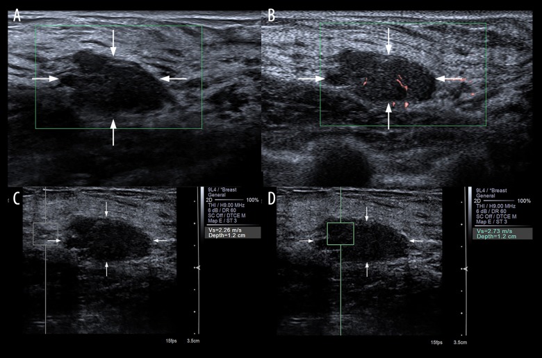
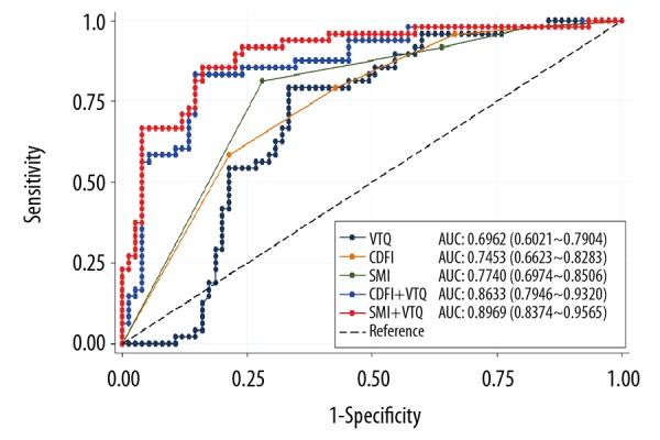
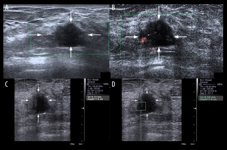
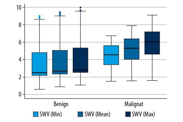
BACKGROUND This study explored the diagnostic value of a combined modality of Superb Microvascular Imaging (SMI) and shear-wave elastography in differentiating malignant and benign breast lesions. MATERIAL AND METHODS A total of 121 patients with 123 breast lesions enrolled underwent conventional ultrasound exam (US), Color Doppler Flow Imaging (CDFI), SMI examination, and Virtual Touch Tissue Quantification (VTQ) measurement between May 2016 and October 2017. Vessels were detected by both CDFI and SMI in a quantitative manner. The stiffness of all the breast tissues was evaluated by VTQ method. We further assessed the diagnostic performances of CDFI, SMI, VTQ, CDFI+VTQ, and SMI+VTQ. RESULTS Both CDFI and SMI exhibited significant differences between malignant and benign masses (p<0.001) in terms of Adler classification. The mean shear-wave velocity (SWV) of malignant neoplasms was 5.28 m/s, with interquartile range (IQR) 4.01-6.39 m/s (p<0.001). The mean SWV of benign lesions was 2.64 m/s, with IQR 2.30-5.01 m/s (p<0.001). No significant difference was found for the area under the receiver operating characteristic curve (AUC) for CDFI, SMI, and VTQ (c2=2.29, P=0.3715). The sensitivity was the highest on SMI+VTQ (85.42%) and the lowest on CDFI (58.33%). CDFI+VTQ (85.33%) had a slightly higher specificity than SMI+VTQ (84.00%). The accuracy rate of these 2 modalities remained the same (84.55%). CONCLUSIONS Superb Microvascular Imaging yields more detailed vascular information in the bloodstream in benign and malignant breast masses compared with conventional ultrasonography. VTQ provides standardized quantified results in assessing tissue stiffness. The combined modality of SMI+VTQ added to conventional ultrasonography presented a better diagnostic performance in differentiating malignant breast neoplasms.

摘要

背景

本研究探讨了 Superb Microvascular Imaging(SMI)和剪切波弹性成像联合模式在鉴别乳腺良恶性病变中的诊断价值。

材料与方法

2016 年 5 月至 2017 年 10 月期间,共 121 例 123 个乳腺病灶的患者接受了常规超声检查(US)、彩色多普勒血流成像(CDFI)、SMI 检查和虚拟触诊组织定量(VTQ)测量。通过 CDFI 和 SMI 对血管进行定量检测,采用 VTQ 方法评估所有乳腺组织的硬度。我们进一步评估了 CDFI、SMI、VTQ、CDFI+VTQ 和 SMI+VTQ 的诊断性能。

结果

CDFI 和 SMI 在 Adler 分类方面均显示出良恶性肿块之间的显著差异(p<0.001)。恶性肿瘤的平均剪切波速度(SWV)为 5.28 m/s,四分位距(IQR)为 4.01-6.39 m/s(p<0.001)。良性病变的平均 SWV 为 2.64 m/s,IQR 为 2.30-5.01 m/s(p<0.001)。CDFI、SMI 和 VTQ 的受试者工作特征曲线(ROC)下面积(AUC)无显著差异(c2=2.29,P=0.3715)。SMI+VTQ 的敏感性最高(85.42%),CDFI 的敏感性最低(58.33%)。CDFI+VTQ(85.33%)的特异性略高于 SMI+VTQ(84.00%)。这两种模式的准确率相同(84.55%)。

结论

与常规超声相比,SMI 可提供乳腺良恶性肿块中血流更详细的血管信息。VTQ 提供评估组织硬度的标准化定量结果。SMI+VTQ 联合常规超声在鉴别乳腺恶性肿瘤方面具有更好的诊断性能。

https://cdn.ncbi.nlm.nih.gov/pmc/blobs/7952/6122269/9160a63e774d/medscimonit-24-5935-g004.jpg
https://cdn.ncbi.nlm.nih.gov/pmc/blobs/7952/6122269/f21cbb779378/medscimonit-24-5935-g001.jpg
https://cdn.ncbi.nlm.nih.gov/pmc/blobs/7952/6122269/119f6168230a/medscimonit-24-5935-g002.jpg
https://cdn.ncbi.nlm.nih.gov/pmc/blobs/7952/6122269/cdf484ba05ae/medscimonit-24-5935-g003.jpg
https://cdn.ncbi.nlm.nih.gov/pmc/blobs/7952/6122269/9160a63e774d/medscimonit-24-5935-g004.jpg

相似文献

1
Diagnostic Performance of Superb Microvascular Imaging (SMI) Combined with Shear-Wave Elastography in Evaluating Breast Lesions.超声微血管成像(SMI)联合剪切波弹性成像对乳腺病变的诊断性能评估。
Med Sci Monit. 2018 Aug 26;24:5935-5942. doi: 10.12659/MSM.910399.
2
The Diagnostic Value of Superb Microvascular Imaging (SMI) in Detecting Blood Flow Signals of Breast Lesions: A Preliminary Study Comparing SMI to Color Doppler Flow Imaging.超微血管成像(SMI)在检测乳腺病变血流信号中的诊断价值:一项比较SMI与彩色多普勒血流成像的初步研究
Medicine (Baltimore). 2015 Sep;94(36):e1502. doi: 10.1097/MD.0000000000001502.
3
A Prospective Study to Compare Superb Microvascular Imaging with Grayscale Ultrasound and Color Doppler Flow Imaging of Vascular Distribution and Morphology in Thyroid Nodules.一项对比甲状腺结节血管分布和形态的灰阶超声及彩色多普勒血流显像与超级微血管成像的前瞻性研究。
Med Sci Monit. 2018 Dec 19;24:9223-9231. doi: 10.12659/MSM.911695.
4
Role of Superb Microvascular Imaging in Differentiating Between Malignant and Benign Solid Breast Masses.超级微血管成像在鉴别良恶性实性乳腺肿块中的作用。
Clin Breast Cancer. 2020 Dec;20(6):e786-e793. doi: 10.1016/j.clbc.2020.06.009. Epub 2020 Jul 3.
5
[Application value of superb microvascular imaging for diagnosis of different size renal solid tumors].[超微血管成像在不同大小肾实性肿瘤诊断中的应用价值]
Zhonghua Zhong Liu Za Zhi. 2021 Nov 23;43(11):1215-1221. doi: 10.3760/cma.j.cn112152-20191220-00832.
6
Superb microvascular imaging in diagnosis of breast lesions: a comparative study with contrast-enhanced ultrasonographic microvascular imaging.超微血管成像在乳腺病变诊断中的应用:与超声造影微血管成像的对比研究
Br J Radiol. 2016 Oct;89(1066):20160546. doi: 10.1259/bjr.20160546. Epub 2016 Aug 16.
7
The diagnostic performance of shear wave velocity ratio for the differential diagnosis of benign and malignant breast lesions: Compared with VTQ, and mammography.剪切波速度比值对良恶性乳腺病变的鉴别诊断性能:与 VTQ 和钼靶相比。
Clin Hemorheol Microcirc. 2021;77(2):123-131. doi: 10.3233/CH-200813.
8
Combination of Quantitative Parameters of Shear Wave Elastography and Superb Microvascular Imaging to Evaluate Breast Masses.剪切波弹性成像定量参数与超微血管成像联合评估乳腺肿块。
Korean J Radiol. 2020 Sep;21(9):1045-1054. doi: 10.3348/kjr.2019.0765.
9
Superb Microvascular Imaging-A new vascular detecting ultrasonographic technique for avascular breast masses: A preliminary study.超微血管成像——一种用于无血管乳腺肿块的新型血管检测超声技术:一项初步研究。
Eur J Radiol. 2016 May;85(5):915-21. doi: 10.1016/j.ejrad.2015.12.011. Epub 2016 Jan 18.
10
A comparative study on superb microvascular imaging and conventional ultrasonography in differentiating BI-RADS 4 breast lesions.超声微血管成像与传统超声鉴别BI-RADS 4类乳腺病变的对比研究
Oncol Lett. 2019 Sep;18(3):3202-3210. doi: 10.3892/ol.2019.10603. Epub 2019 Jul 11.

引用本文的文献

1
Advanced multimodal ultrasound for pre-operative assessment of skin tumours: A case series.用于皮肤肿瘤术前评估的先进多模态超声:病例系列
Ultrasound. 2024 Nov 30:1742271X241289021. doi: 10.1177/1742271X241289021.
2
Evaluating the stage of deep vein thrombosis: the diagnostic accuracy of shear wave elastography and super-microvascular imaging.评估深静脉血栓形成的阶段:剪切波弹性成像和超微血管成像的诊断准确性。
Quant Imaging Med Surg. 2024 Sep 1;14(9):6882-6894. doi: 10.21037/qims-23-1822. Epub 2024 May 21.
3
Diagnostic value of contrast-enhanced ultrasound and shear-wave elastography for small breast nodules.

本文引用的文献

1
Benefit of Shear-wave Elastography in the differential diagnosis of breast lesion: a diagnostic meta-analysis.剪切波弹性成像在乳腺病变鉴别诊断中的益处:一项诊断性荟萃分析。
Med Ultrason. 2018 Feb 4;1(1):43-49. doi: 10.11152/mu-1209.
2
Application of superb microvascular imaging in focal liver lesions.超微血管成像在肝脏局灶性病变中的应用。
World J Gastroenterol. 2017 Nov 21;23(43):7765-7775. doi: 10.3748/wjg.v23.i43.7765.
3
Breast lesions classified as probably benign (BI-RADS 3) on magnetic resonance imaging: a systematic review and meta-analysis.
超声造影及剪切波弹性成像对乳腺小结节的诊断价值
PeerJ. 2024 Jul 3;12:e17677. doi: 10.7717/peerj.17677. eCollection 2024.
4
Using a combination of superb microvascular imaging and other auxiliary ultrasound techniques to increase the accuracy of gray-scale ultrasound for breast masses.采用卓越的微血管成像技术和其他辅助超声技术相结合的方法,提高乳腺肿块灰阶超声的准确性。
BMC Cancer. 2024 Feb 16;24(1):224. doi: 10.1186/s12885-024-11981-9.
5
Multimodal ultrasound imaging for diagnostic differentiation of sclerosing adenosis from invasive ductal carcinoma.多模态超声成像用于硬化性腺病与浸润性导管癌的诊断鉴别
Quant Imaging Med Surg. 2024 Jan 3;14(1):877-887. doi: 10.21037/qims-23-524. Epub 2024 Jan 2.
6
Recent Advances in Ultrasound Breast Imaging: From Industry to Clinical Practice.超声乳腺成像的最新进展:从工业到临床实践
Diagnostics (Basel). 2023 Mar 4;13(5):980. doi: 10.3390/diagnostics13050980.
7
Development and internal validation of a conventional ultrasound-based nomogram for predicting malignant nonmasslike breast lesions.用于预测乳腺非肿块样恶性病变的传统超声列线图的开发与内部验证
Quant Imaging Med Surg. 2022 Dec;12(12):5452-5461. doi: 10.21037/qims-22-378.
8
Diagnostic accuracy of ultrasound shear wave elastography combined with superb microvascular imaging for breast tumors: A protocol for systematic review and meta-analysis.超声剪切波弹性成像联合超微血管成像对乳腺肿瘤的诊断准确性:系统评价与Meta分析方案
Medicine (Baltimore). 2021 Jun 25;100(25):e26262. doi: 10.1097/MD.0000000000026262.
9
Assessment of neovascularization of carotid artery atherosclerotic plaques using superb microvascular imaging: a comparison with contrast-enhanced ultrasound imaging and histology.使用超微血管成像评估颈动脉粥样硬化斑块的新生血管形成:与对比增强超声成像和组织学的比较。
Quant Imaging Med Surg. 2021 May;11(5):1958-1969. doi: 10.21037/qims-20-933.
10
Evaluation of Multimode Color Doppler Flow Imaging in the Diagnosis of Solid Renal Tumor.多模态彩色多普勒血流成像在诊断肾脏实性肿瘤中的评价
Contrast Media Mol Imaging. 2021 Apr 1;2021:6656877. doi: 10.1155/2021/6656877. eCollection 2021.
磁共振成像中分类为可能良性(BI-RADS 3)的乳腺病变:系统评价和荟萃分析。
Eur Radiol. 2018 May;28(5):1919-1928. doi: 10.1007/s00330-017-5127-y. Epub 2017 Nov 22.
4
The value of superb microvascular imaging in differentiating benign renal mass from malignant renal tumor: a retrospective study.超微血管成像在鉴别肾良性肿块与肾恶性肿瘤中的价值:一项回顾性研究。
Br J Radiol. 2018 Feb;91(1082):20170601. doi: 10.1259/bjr.20170601. Epub 2017 Nov 28.
5
Three-dimensional versus two-dimensional shear-wave elastography: Associations of mean elasticity values with prognostic factors and tumor subtypes of breast cancer.三维与二维剪切波弹性成像:平均弹性值与乳腺癌预后因素及肿瘤亚型的相关性
Clin Imaging. 2018 Mar-Apr;48:79-85. doi: 10.1016/j.clinimag.2017.10.007. Epub 2017 Oct 12.
6
Value of shear wave arrival time contour display in shear wave elastography for breast masses diagnosis.剪切波到达时间轮廓显示在剪切波弹性成像中的价值用于乳腺肿块的诊断。
Sci Rep. 2017 Aug 1;7(1):7036. doi: 10.1038/s41598-017-07389-0.
7
Preliminary Evaluation of Virtual Touch Tissue Imaging Quantification for Differential Diagnosis of Metastatic and Nonmetastatic Cervical Lymph Nodes.虚拟触诊组织成像定量技术在转移性与非转移性宫颈淋巴结鉴别诊断中的初步评估
J Ultrasound Med. 2017 Mar;36(3):557-563. doi: 10.7863/ultra.16.03077. Epub 2017 Jan 27.
8
Descriptive epidemiology of breast cancer in China: incidence, mortality, survival and prevalence.中国乳腺癌的描述性流行病学:发病率、死亡率、生存率和患病率。
Breast Cancer Res Treat. 2016 Oct;159(3):395-406. doi: 10.1007/s10549-016-3947-0. Epub 2016 Aug 25.
9
Superb microvascular imaging in diagnosis of breast lesions: a comparative study with contrast-enhanced ultrasonographic microvascular imaging.超微血管成像在乳腺病变诊断中的应用:与超声造影微血管成像的对比研究
Br J Radiol. 2016 Oct;89(1066):20160546. doi: 10.1259/bjr.20160546. Epub 2016 Aug 16.
10
Comparison of the superb microvascular imaging technique and the color Doppler techniques for evaluating children's testicular blood flow.用于评估儿童睾丸血流的超微血管成像技术与彩色多普勒技术的比较。
Eur Rev Med Pharmacol Sci. 2016 May;20(10):1947-53.