Faculty of Life and Environmental Sciences, University of Tsukuba, Tsukuba, Japan
Microbiology Research Center for Sustainability, University of Tsukuba, Tsukuba, Japan.
Appl Environ Microbiol. 2019 Aug 29;85(18). doi: 10.1128/AEM.00608-19. Print 2019 Sep 15.
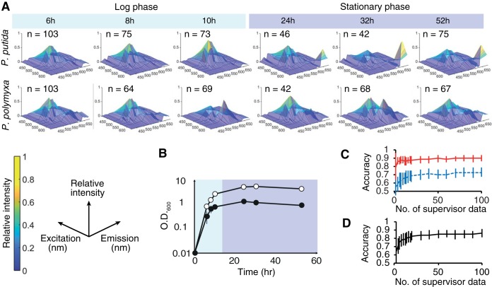
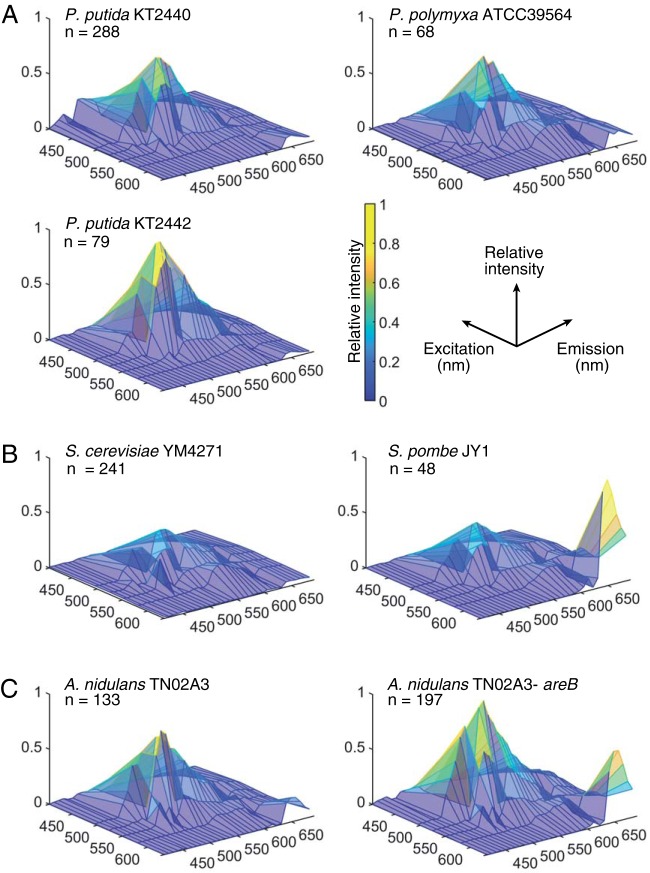
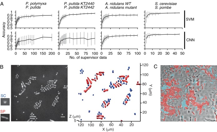
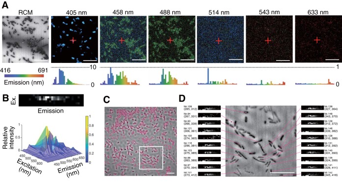
Here we analyzed the innate fluorescence signature of the single microbial cell, within both clonal and mixed populations of microorganisms. We found that even very similarly shaped cells differ noticeably in their autofluorescence features and that the innate fluorescence signatures change dynamically with growth phases. We demonstrated that machine learning models can be trained with a data set of single-cell innate fluorescence signatures to annotate cells according to their phenotypes and physiological status, for example, distinguishing a wild-type cell from its nitrogen metabolism mutant counterpart and log-phase cells from stationary-phase cells of We developed a minimally invasive method (onfocal eflection microscopy-assisted single-cell nnate luorescence [CRIF] analysis) to optically extract and catalog the innate cellular fluorescence signatures of each of the individual live microbial cells in a three-dimensional space. This technique represents a step forward from traditional techniques which analyze the innate fluorescence signatures at the population level and necessitate a clonal culture. Since the fluorescence signature is an innate property of a cell, our technique allows the prediction of the types or physiological status of intact and tag-free single cells, within a cell population distributed in a three-dimensional space. Our study presents a blueprint for a streamlined cell analysis where one can directly assess the potential phenotype of each single cell in a heterogenous population by its autofluorescence signature under a microscope, without cell tagging. A cell's innate fluorescence signature is an assemblage of fluorescence signals emitted by diverse biomolecules within a cell. It is known that the innate fluoresce signature reflects various cellular properties and physiological statuses; thus, they can serve as a rich source of information in cell characterization as well as cell identification. However, conventional techniques focus on the analysis of the innate fluorescence signatures at the population level but not at the single-cell level and thus necessitate a clonal culture. In the present study, we developed a technique to analyze the innate fluorescence signature of a single microbial cell. Using this novel method, we found that even very similarly shaped cells differ noticeably in their autofluorescence features, and the innate fluorescence signature changes dynamically with growth phases. We also demonstrated that the different cell types can be classified accurately within a mixed population under a microscope at the resolution of a single cell, depending solely on the innate fluorescence signature information. We suggest that single-cell autofluoresce signature analysis is a promising tool to directly assess the taxonomic or physiological heterogeneity within a microbial population, without cell tagging.
在这里,我们分析了单个微生物细胞的固有荧光特征,包括微生物的克隆和混合群体。我们发现,即使形状非常相似的细胞,其自发荧光特征也有明显差异,并且固有荧光特征随生长阶段而动态变化。我们证明,可以使用单细胞固有荧光特征的数据集来训练机器学习模型,根据细胞的表型和生理状态对细胞进行注释,例如,将野生型细胞与氮代谢突变体细胞区分开来,将对数期细胞与静止期细胞区分开来。我们开发了一种微创方法(共聚焦反射显微镜辅助单细胞固有荧光[CRIF]分析),以光学方式提取和编目每个活微生物细胞在三维空间中的固有细胞荧光特征。与需要克隆培养的传统技术相比,该技术代表了向前迈出的一步,因为传统技术仅在群体水平上分析固有荧光特征。由于荧光特征是细胞的固有特性,因此我们的技术允许在三维空间中分布的细胞群体中,对完整且无标记的单个细胞的类型或生理状态进行预测。我们的研究为简化的细胞分析提供了蓝图,可以在显微镜下通过细胞的自发荧光特征直接评估异质群体中每个单个细胞的潜在表型,而无需对细胞进行标记。细胞的固有荧光特征是细胞内各种生物分子发出的荧光信号的组合。已知固有荧光特征反映了各种细胞特性和生理状态;因此,它们可以作为细胞特征描述以及细胞识别的丰富信息来源。然而,传统技术侧重于群体水平而不是单细胞水平的固有荧光特征分析,因此需要克隆培养。在本研究中,我们开发了一种分析单个微生物细胞固有荧光特征的技术。使用这种新方法,我们发现即使形状非常相似的细胞,其自发荧光特征也有明显差异,并且固有荧光特征随生长阶段而动态变化。我们还证明,在显微镜下,仅根据固有荧光特征信息,就可以在混合群体中准确地对不同类型的细胞进行分类,分类分辨率可达单个细胞。我们认为,单细胞自发荧光特征分析是一种很有前途的工具,可以直接评估微生物群体中的分类或生理异质性,而无需对细胞进行标记。