• 文献检索
  • 文档翻译
  • 深度研究
  • 学术资讯
  • Suppr Zotero 插件Zotero 插件
  • 邀请有礼
  • 套餐&价格
  • 历史记录
应用&插件
Suppr Zotero 插件Zotero 插件浏览器插件Mac 客户端Windows 客户端微信小程序
定价
高级版会员购买积分包购买API积分包
服务
文献检索文档翻译深度研究API 文档MCP 服务
关于我们
关于 Suppr公司介绍联系我们用户协议隐私条款
关注我们

Suppr 超能文献

核心技术专利:CN118964589B侵权必究
粤ICP备2023148730 号-1Suppr @ 2026

文献检索

告别复杂PubMed语法,用中文像聊天一样搜索,搜遍4000万医学文献。AI智能推荐,让科研检索更轻松。

立即免费搜索

文件翻译

保留排版,准确专业,支持PDF/Word/PPT等文件格式,支持 12+语言互译。

免费翻译文档

深度研究

AI帮你快速写综述,25分钟生成高质量综述,智能提取关键信息,辅助科研写作。

立即免费体验

基于 CT 的肿瘤密实度预测食管鳞癌多模态治疗后的预后。

Tumor Compactness based on CT to predict prognosis after multimodal treatment for esophageal squamous cell carcinoma.

机构信息

Department of Radiation Oncology, Sichuan Cancer Hospital & Institution, Sichuan Cancer Center, School of Medicine, University of Electronic Science and Technology of China, Radiation oncology Key Laboratory of Sichuan Province, Chengdu, 610041, China.

Department of Radiation Oncology, Fujian Cancer Hospital & Fujian Medical University Cancer Hospital, Fuzhou, 350014, China.

出版信息

Sci Rep. 2019 Jul 19;9(1):10497. doi: 10.1038/s41598-019-46899-x.

DOI:10.1038/s41598-019-46899-x
PMID:31324827
原文链接:https://pmc.ncbi.nlm.nih.gov/articles/PMC6642095/
Abstract

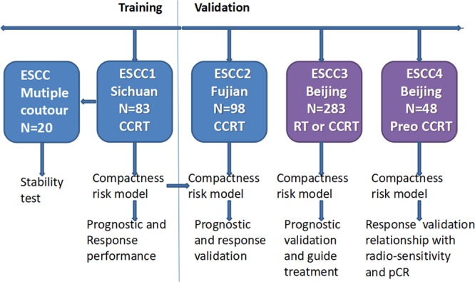
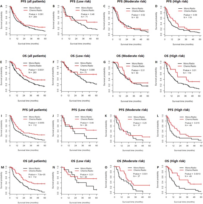
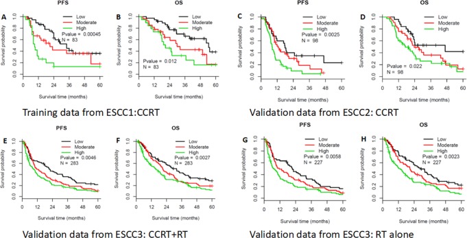
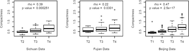
We aimed to establish a risk model using computed tomography-based compactness to predict overall survival (OS) and progression-free survival (PFS) after multimodal treatment for esophageal squamous cell carcinoma (ESCC). We extracted pre-treatment computed tomography-based tumor data (volume, surface area, and compactness) for 512 cases of ESCC that were treated at 3 centers. A risk model based on compactness was trained using Cox regression analyses of data from 83 cases, and then the model was validated using two independent cohorts (98 patients and 283 patients). The largest cohort (283 patients) was then evaluated using the risk model to predict response to radiotherapy with or without chemotherapy. In the three datasets, the pre-treatment compactness risk model provided good accuracy for predicting OS (P = 0.012, P = 0.022, and P = 0.003) and PFS (P < 0.001, P = 0.003, and P = 0.005). Patients in the low-risk group did not experience a significant OS benefit from concurrent chemoradiotherapy (P = 0.099). Furthermore, after preoperative concurrent chemoradiotherapy, the OS outcomes were similar among patients in the low-risk group who did and did not achieve a pathological complete response (P = 0.127). Tumor compactness was correlated with clinical T stage but was more accurate for predicting prognosis after treatment for ESCC, based on higher C-index values in all three datasets. The compactness-based risk model was effective for predicting OS and PFS after multimodal treatment for ESCC. Therefore, it may be useful for guiding personalized treatment.

摘要

我们旨在建立一个基于 CT 紧凑度的风险模型,以预测食管鳞癌(ESCC)经多模态治疗后的总生存期(OS)和无进展生存期(PFS)。我们从 3 个中心提取了 512 例 ESCC 患者的治疗前 CT 肿瘤数据(体积、表面积和紧凑度)。使用来自 83 例数据的 Cox 回归分析,基于紧凑度建立风险模型,然后使用两个独立队列(98 例患者和 283 例患者)验证模型。使用最大队列(283 例患者)评估风险模型,以预测放疗联合或不联合化疗的反应。在这三个数据集,治疗前的紧凑度风险模型对于预测 OS(P=0.012,P=0.022,P=0.003)和 PFS(P<0.001,P=0.003,P=0.005)具有良好的准确性。低风险组患者在同步放化疗中并未显著获益(P=0.099)。此外,在术前同步放化疗后,低风险组中达到和未达到病理完全缓解的患者的 OS 结果相似(P=0.127)。肿瘤紧凑度与临床 T 分期相关,但基于所有三个数据集更高的 C 指数值,对于预测 ESCC 治疗后预后更准确。基于 CT 紧凑度的风险模型可有效预测 ESCC 经多模态治疗后的 OS 和 PFS。因此,它可能有助于指导个性化治疗。

https://cdn.ncbi.nlm.nih.gov/pmc/blobs/2ede/6642095/a88696fc0bff/41598_2019_46899_Fig4_HTML.jpg
https://cdn.ncbi.nlm.nih.gov/pmc/blobs/2ede/6642095/8ff5e7ce9e4a/41598_2019_46899_Fig1_HTML.jpg
https://cdn.ncbi.nlm.nih.gov/pmc/blobs/2ede/6642095/712323b301af/41598_2019_46899_Fig2_HTML.jpg
https://cdn.ncbi.nlm.nih.gov/pmc/blobs/2ede/6642095/00d1d5b2e445/41598_2019_46899_Fig3_HTML.jpg
https://cdn.ncbi.nlm.nih.gov/pmc/blobs/2ede/6642095/a88696fc0bff/41598_2019_46899_Fig4_HTML.jpg

相似文献

1
Tumor Compactness based on CT to predict prognosis after multimodal treatment for esophageal squamous cell carcinoma.基于 CT 的肿瘤密实度预测食管鳞癌多模态治疗后的预后。
Sci Rep. 2019 Jul 19;9(1):10497. doi: 10.1038/s41598-019-46899-x.
2
Prediction of the tumor response and survival based on computed tomography in esophageal squamous cell carcinoma after trimodality therapy.基于三模态治疗后食管鳞癌 CT 预测肿瘤反应和生存。
Surg Today. 2021 Sep;51(9):1496-1505. doi: 10.1007/s00595-021-02277-7. Epub 2021 Apr 2.
3
Combined modalities of magnetic resonance imaging, endoscopy and computed tomography in the evaluation of tumor responses to definitive chemoradiotherapy in esophageal squamous cell carcinoma.磁共振成像、内窥镜检查和计算机断层扫描联合模式在评估食管鳞状细胞癌根治性放化疗后的肿瘤反应中的应用
Radiother Oncol. 2016 Nov;121(2):239-245. doi: 10.1016/j.radonc.2016.09.017. Epub 2016 Oct 25.
4
Application of sequential (18)F-FDG PET/CT scans for concurrent chemoradiotherapy of non-surgical squamous cell esophageal carcinoma.序贯(18)F-FDG PET/CT扫描在非手术治疗的食管鳞状细胞癌同步放化疗中的应用
J BUON. 2014 Apr-Jun;19(2):517-23.
5
Proposed revision of the 8th edition AJCC clinical staging system for esophageal squamous cell cancer treated with definitive chemo-IMRT based on CT imaging.基于 CT 影像学的根治性放化疗治疗食管鳞癌第 8 版 AJCC 临床分期系统的修订建议。
Radiat Oncol. 2019 Mar 28;14(1):54. doi: 10.1186/s13014-019-1258-4.
6
Pre-treatment maximal oesophageal wall thickness is independently associated with response to chemoradiotherapy in patients with T3-4 oesophageal squamous cell carcinoma.术前最大食管壁厚度与 T3-4 期食管鳞癌患者放化疗反应独立相关。
Eur J Cardiothorac Surg. 2012 Dec;42(6):958-64. doi: 10.1093/ejcts/ezs136. Epub 2012 Mar 30.
7
Tenascin-C, a Prognostic Determinant of Esophageal Squamous Cell Carcinoma.腱生蛋白-C,食管鳞状细胞癌的一个预后决定因素。
PLoS One. 2016 Jan 5;11(1):e0145807. doi: 10.1371/journal.pone.0145807. eCollection 2016.
8
Response evaluation with endoscopic ultrasound and computed tomography in esophageal squamous cell carcinoma treated by definitive chemoradiotherapy.内镜超声和计算机断层扫描在接受根治性放化疗的食管鳞状细胞癌中的疗效评估
J Gastroenterol Hepatol. 2015 Mar;30(3):463-9. doi: 10.1111/jgh.12683.
9
Correlation of plasma miR-21 and miR-93 with radiotherapy and chemotherapy efficacy and prognosis in patients with esophageal squamous cell carcinoma.血浆 miR-21 和 miR-93 与食管鳞癌患者放化疗疗效及预后的相关性。
World J Gastroenterol. 2019 Oct 7;25(37):5604-5618. doi: 10.3748/wjg.v25.i37.5604.
10
Improved prognosis with induction chemotherapy in pathological complete responders after trimodality treatment for esophageal squamous cell carcinoma: Hypothesis generating for adjuvant treatment.新辅助化疗在三模态治疗后病理完全缓解的食管鳞癌患者中改善预后:辅助治疗的假设产生。
Eur J Surg Oncol. 2019 Aug;45(8):1498-1504. doi: 10.1016/j.ejso.2019.03.020. Epub 2019 Mar 19.

引用本文的文献

1
Superhydrophobic Array Devices for the Enhanced Formation of 3D Cancer Models.用于增强 3D 癌症模型形成的超疏水阵列器件。
ACS Nano. 2024 Aug 27;18(34):23637-23654. doi: 10.1021/acsnano.4c08132. Epub 2024 Aug 16.
2
Shape phenotype of thigh fat and muscle and risk of major adverse cardiovascular events after fragility hip fracture.大腿脂肪和肌肉的形态表型与脆性髋部骨折后主要不良心血管事件的风险。
J Cachexia Sarcopenia Muscle. 2024 Feb;15(1):331-341. doi: 10.1002/jcsm.13407. Epub 2023 Dec 21.
3
Prognostic significance and postoperative chemoradiotherapy guiding value of mean platelet volume for locally advanced esophageal squamous cell carcinoma patients.

本文引用的文献

1
Feasibility and efficiency of concurrent chemoradiotherapy with capecitabine and cisplatin versus radiotherapy alone for elderly patients with locally advanced esophageal squamous cell carcinoma: Experience of two centers.卡培他滨和顺铂同期放化疗与单纯放疗治疗局部晚期老年食管鳞癌的可行性和效率:两个中心的经验。
Thorac Cancer. 2018 Jan;9(1):59-65. doi: 10.1111/1759-7714.12536. Epub 2017 Oct 12.
2
Intensity-modulated versus 3-dimensional conformal radiotherapy in the definitive treatment of esophageal cancer: comparison of outcomes and acute toxicity.调强放疗与三维适形放疗在食管癌根治性治疗中的比较:疗效与急性毒性对比
Radiat Oncol. 2017 Aug 15;12(1):131. doi: 10.1186/s13014-017-0863-3.
3
平均血小板体积对局部晚期食管鳞状细胞癌患者的预后意义及术后放化疗指导价值
Front Oncol. 2023 Apr 26;13:1094040. doi: 10.3389/fonc.2023.1094040. eCollection 2023.
4
Methodological quality of machine learning-based quantitative imaging analysis studies in esophageal cancer: a systematic review of clinical outcome prediction after concurrent chemoradiotherapy.基于机器学习的食管癌定量成像分析研究的方法学质量:同步放化疗后临床结局预测的系统评价。
Eur J Nucl Med Mol Imaging. 2022 Jul;49(8):2462-2481. doi: 10.1007/s00259-021-05658-9. Epub 2021 Dec 23.
5
A FDG-PET radiomics signature detects esophageal squamous cell carcinoma patients who do not benefit from chemoradiation.基于 FDG-PET 影像组学特征的签名可预测不能从放化疗中获益的食管鳞癌患者。
Sci Rep. 2020 Oct 19;10(1):17671. doi: 10.1038/s41598-020-74701-w.
Prospective comparison of MR with diffusion-weighted imaging, endoscopic ultrasound, MDCT and positron emission tomography-CT in the pre-operative staging of oesophageal cancer: results from a pilot study.
磁共振成像与扩散加权成像、内镜超声、多层螺旋CT及正电子发射断层扫描-CT在食管癌术前分期中的前瞻性比较:一项初步研究结果
Br J Radiol. 2016 Dec;89(1068):20160087. doi: 10.1259/bjr.20160087. Epub 2016 Oct 21.
4
Gross tumor volume is the prognostic factor for squamous cell esophageal cancer patients treated with definitive radiotherapy.肿瘤总体积是接受根治性放疗的食管鳞状细胞癌患者的预后因素。
J Thorac Dis. 2016 Jun;8(6):1155-61. doi: 10.21037/jtd.2016.04.08.
5
Esophageal cancer statistics in China, 2011: Estimates based on 177 cancer registries.中国食管癌统计数据,2011 年:基于 177 个癌症登记处的估计。
Thorac Cancer. 2016 Mar;7(2):232-7. doi: 10.1111/1759-7714.12322. Epub 2015 Dec 7.
6
Cancer statistics in China, 2015.《中国癌症统计数据 2015》
CA Cancer J Clin. 2016 Mar-Apr;66(2):115-32. doi: 10.3322/caac.21338. Epub 2016 Jan 25.
7
Prognosis of esophageal squamous cell carcinoma patients with preoperative radiotherapy: Comparison of different cancer staging systems.术前放疗的食管鳞癌患者预后:不同癌症分期系统的比较。
Thorac Cancer. 2014 May;5(3):204-10. doi: 10.1111/1759-7714.12079. Epub 2014 Apr 22.
8
Residual lymph node status is an independent prognostic factor in esophageal squamous cell Carcinoma with pathologic T0 after preoperative radiotherapy.残留淋巴结状态是术前放疗后病理T0期食管鳞状细胞癌的独立预后因素。
Radiat Oncol. 2015 Jul 11;10:142. doi: 10.1186/s13014-015-0450-4.
9
IBEX: an open infrastructure software platform to facilitate collaborative work in radiomics.IBEX:一个促进放射组学协作工作的开放式基础设施软件平台。
Med Phys. 2015 Mar;42(3):1341-53. doi: 10.1118/1.4908210.
10
Decoding tumour phenotype by noninvasive imaging using a quantitative radiomics approach.采用定量放射组学方法通过无创成像解码肿瘤表型。
Nat Commun. 2014 Jun 3;5:4006. doi: 10.1038/ncomms5006.