Yakovliev A, Ziniuk R, Wang D, Xue B, Vretik L O, Nikolaeva O A, Tan M, Chen G, Slominskii Yu L, Qu J, Ohulchanskyy T Y
Key Laboratory of Optoelectronic Devices and Systems of Ministry of Education and Guangdong Province, College of Physics and Optoelectronic Engineering, Shenzhen University, Shenzhen, Guangdong Province, 518060, People's Republic of China.
Taras Shevchenko National University of Kyiv, Kyiv, 01601, Ukraine.
Nanoscale Res Lett. 2019 Jul 19;14(1):243. doi: 10.1186/s11671-019-3068-x.
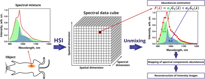
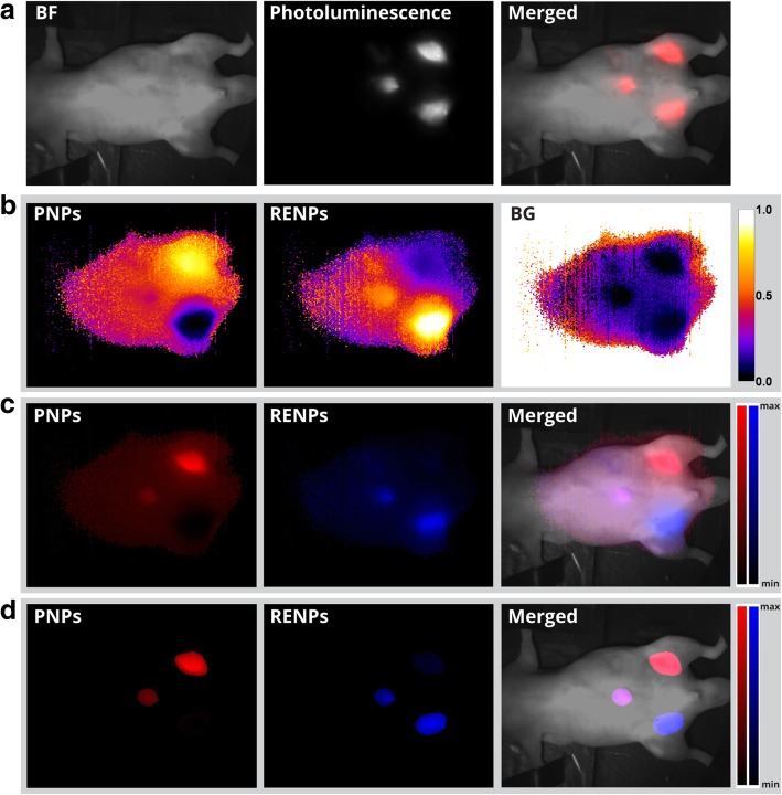
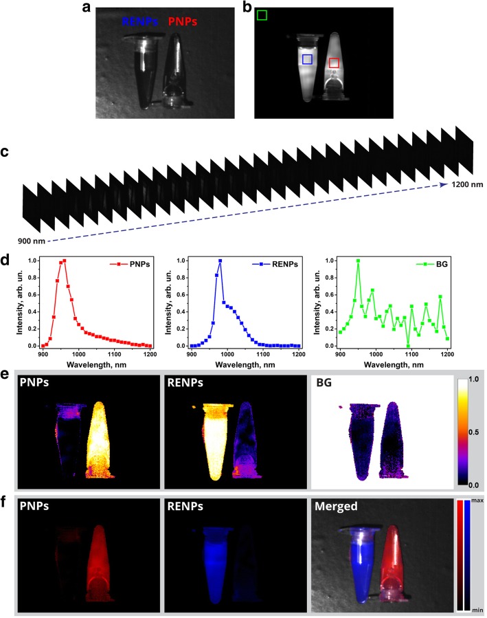
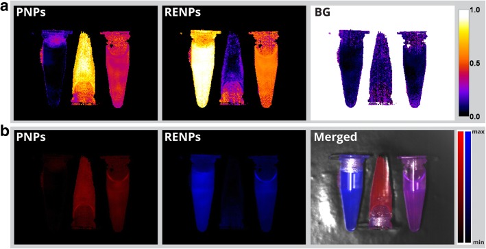
Optical bioimaging with exogenous luminophores emitting in short-wave infrared spectral region (SWIR, ~ 1000-1700 nm) is a rapidly developing field, and the development of multiple SWIR-photoluminescent nanoprobes has recently been reported. In this regard, hyperspectral imaging (HSI), combined with unmixing algorithms, is a promising tool that can allow for efficient multiplexing of the SWIR-emitting nanoagents by their photoluminescence (PL) spectral profiles. The SWIR HSI technique reported here is developed to multiplex two types of nanoprobes: polymeric nanoparticles doped with organic dye (PNPs) and rare-earth doped fluoride nanoparticles (RENPs). Both types of nanoprobes exhibit PL in the same spectral range (~ 900-1200 nm), which hinders spectral separation of PL with optical filters and limits possibilities for their multiplexed imaging in biological tissues. By applying SWIR HSI, we exploited differences in the PL spectral profiles and achieved the spectrally selective and sensitive imaging of the PL signal from every type of nanoparticles. Unmixing of acquired data allowed for multiplexing of the spectrally overlapping nanoprobes by their PL profile. Both quantitative and spatial distribution for every type of nanoparticles were obtained from their mixed suspensions. Finally, the SWIR HSI technique with unmixing protocol was applied to in vivo imaging of mice subcutaneously injected with PNPs and RENPs. The applicability of hyperspectral techniques to multiplex nanoprobes in the in vivo imaging was successfully demonstrated.
利用在短波红外光谱区域(SWIR,1000 - 1700 nm)发射的外源性发光体进行光学生物成像,是一个快速发展的领域,最近已有多种短波红外光致发光纳米探针的相关报道。在这方面,结合解混算法的高光谱成像(HSI)是一种很有前景的工具,它可以通过光致发光(PL)光谱轮廓对发射短波红外光的纳米试剂进行高效复用。本文报道的短波红外高光谱成像技术旨在对两种类型的纳米探针进行复用:掺杂有机染料的聚合物纳米颗粒(PNP)和稀土掺杂氟化物纳米颗粒(RENPs)。这两种类型的纳米探针在相同光谱范围(900 - 1200 nm)内都表现出光致发光,这阻碍了用光学滤光片对光致发光进行光谱分离,并限制了它们在生物组织中进行复用成像的可能性。通过应用短波红外高光谱成像,我们利用了光致发光光谱轮廓的差异,实现了对每种类型纳米颗粒的光致发光信号进行光谱选择性和灵敏成像。对采集数据的解混使得能够通过光谱重叠的纳米探针的光致发光轮廓进行复用。从它们的混合悬浮液中获得了每种类型纳米颗粒的定量和空间分布。最后,将具有解混协议的短波红外高光谱成像技术应用于皮下注射了PNP和RENPs的小鼠的体内成像。成功证明了高光谱技术在体内成像中对纳米探针复用的适用性。