Department of Experimental and Clinical Biomedical Sciences "Mario Serio", University of Florence, Florence, Italy.
Excellence Centre for Research, Transfer and High Education for the development of DE NOVO Therapies (DENOTHE), Florence, Italy.
Nephrol Dial Transplant. 2021 Jan 1;36(1):19-28. doi: 10.1093/ndt/gfz136.
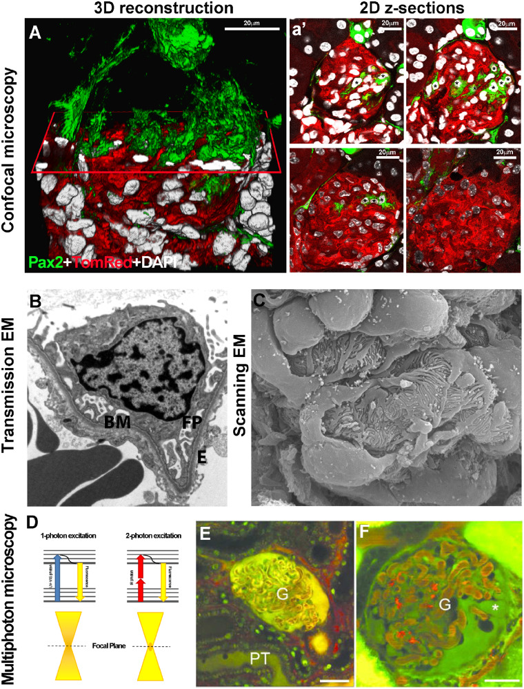
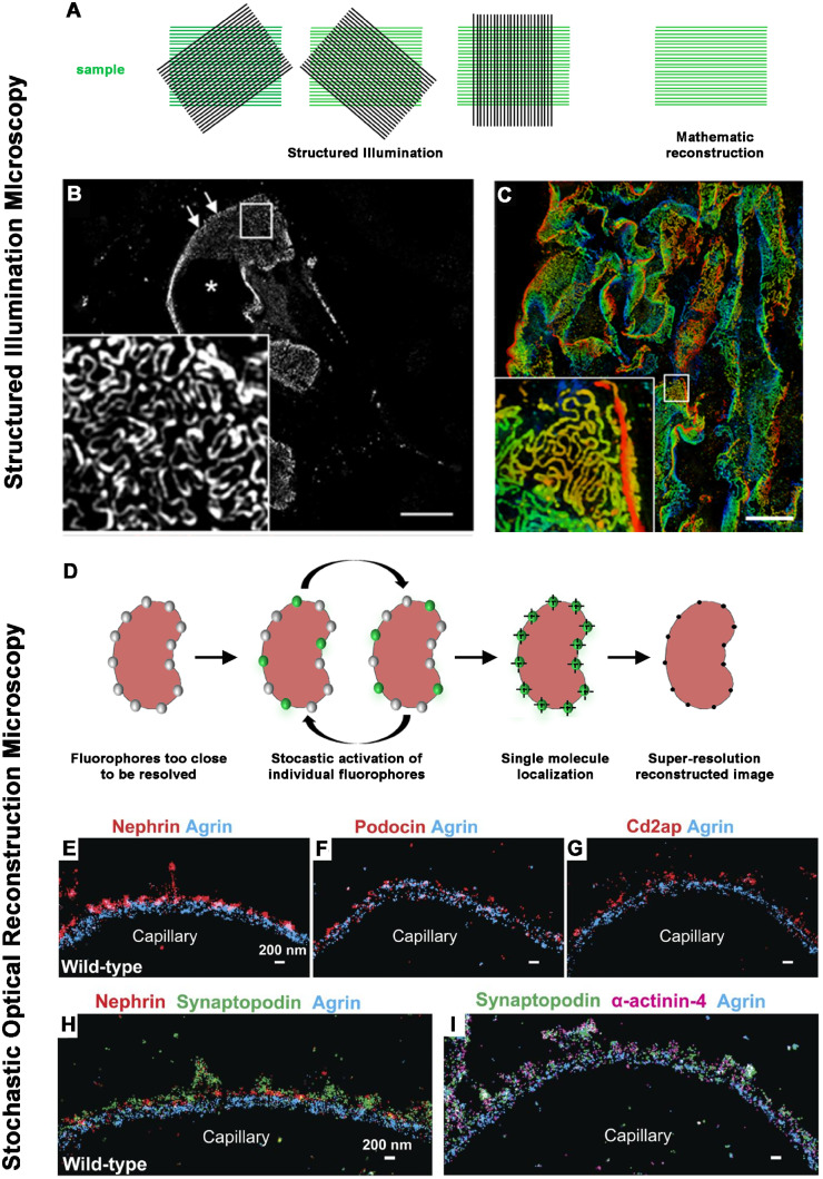
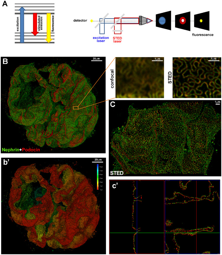
The important achievements in kidney physiological and pathophysiological mechanisms can largely be ascribed to progress in the technology of microscopy. Much of what we know about the architecture of the kidney is based on the fundamental descriptions of anatomic microscopists using light microscopy and later by ultrastructural analysis provided by electron microscopy. These two techniques were used for the first classification systems of kidney diseases and for their constant updates. More recently, a series of novel imaging techniques added the analysis in further dimensions of time and space. Confocal microscopy allowed us to sequentially visualize optical sections along the z-axis and the availability of specific analysis software provided a three-dimensional rendering of thicker tissue specimens. Multiphoton microscopy permitted us to simultaneously investigate kidney function and structure in real time. Fluorescence-lifetime imaging microscopy allowed to study the spatial distribution of metabolites. Super-resolution microscopy increased sensitivity and resolution up to nanoscale levels. With cryo-electron microscopy, researchers could visualize the individual biomolecules at atomic levels directly in the tissues and understand their interaction at subcellular levels. Finally, matrix-assisted laser desorption/ionization imaging mass spectrometry permitted the measuring of hundreds of different molecules at the same time on tissue sections at high resolution. This review provides an overview of available kidney imaging strategies, with a focus on the possible impact of the most recent technical improvements.
肾脏生理和病理生理机制的重要成果在很大程度上归因于显微镜技术的进步。我们对肾脏结构的了解很大程度上基于光镜下的解剖显微镜学家的基本描述,后来则通过电子显微镜提供的超微结构分析。这两种技术最初用于肾脏疾病的分类系统,并不断进行更新。最近,一系列新的成像技术在时间和空间的更多维度上增加了分析。共聚焦显微镜使我们能够沿 z 轴顺序可视化光学切片,而专用分析软件的可用性则为较厚组织标本提供了三维渲染。多光子显微镜允许我们实时同时研究肾脏功能和结构。荧光寿命成像显微镜允许研究代谢物的空间分布。超分辨率显微镜将灵敏度和分辨率提高到纳米级水平。通过冷冻电子显微镜,研究人员可以直接在组织中观察到单个生物分子的原子水平,并了解它们在亚细胞水平的相互作用。最后,基质辅助激光解吸/电离成像质谱允许在高分辨率的组织切片上同时测量数百种不同的分子。这篇综述提供了可用的肾脏成像策略概述,重点介绍了最近技术改进的可能影响。