Edrees Hadeel Y, Abu Zeid Sawsan T H, Atta Hazem M, AlQriqri Mehal A
Endodontic Department, Faculty of Dentistry, King Abdulaziz University, Jeddah 22252, Saudi Arabia.
Endodontic Department, Faculty of Oral and Dental Medicine, Cairo University, Cairo 12345, Egypt.
Materials (Basel). 2019 Jul 19;12(14):2311. doi: 10.3390/ma12142311.
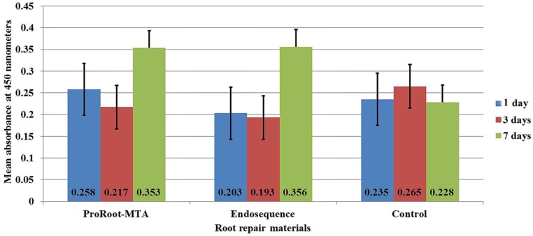
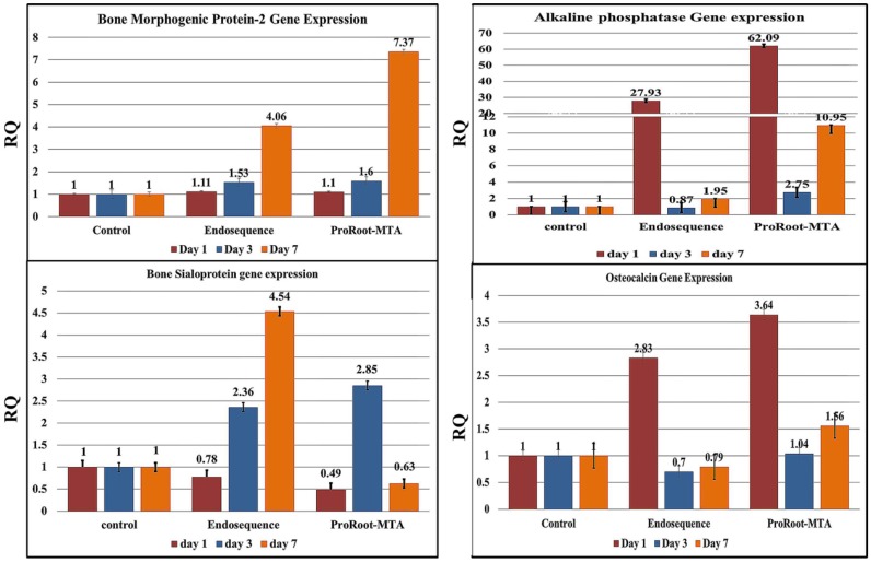
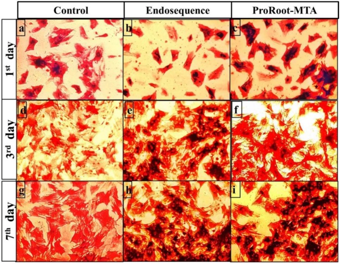
This study aimed to evaluate the osteogenic activity of Endosequence Root Repair Material (ERRM) putty using rat mesenchymal stem cells (MSCs). The extract of set ERRM and ProRoot-mineral trioxide aggregate (MTA) (control) was cocultured with rat MSCs and incubated for one, three, and seven days. The cell viability and proliferation were assessed. A quantitative real-time polymerase chain reaction for bone morphogenetic protein-2 (BMP-2), alkaline phosphatase, bone sialoprotein, and osteocalcin gene expression was performed. Both materials enhanced cell viability and proliferation, which increased over time. On day seven, the cells treated with either material exhibited significantly greater cell viability compared with control untreated cells. MSCs treated with either material showed deeper alkaline phosphatase staining after three days compared to control untreated cells. Treated MSCs also exhibited upregulation of the gene expression of bone morphogenetic protein-2, alkaline phosphatase, bone sialoprotein, and osteocalcin. Both ERRM and ProRoot-MTA enhance the osteogenic differentiation of MSCs.
本研究旨在使用大鼠间充质干细胞(MSCs)评估Endosequence根修复材料(ERRM)糊剂的成骨活性。将凝固后的ERRM和ProRoot-矿物三氧化物凝聚体(MTA)(对照)提取物与大鼠MSCs共培养,并孵育1天、3天和7天。评估细胞活力和增殖情况。进行了骨形态发生蛋白-2(BMP-2)、碱性磷酸酶、骨唾液蛋白和骨钙素基因表达的定量实时聚合酶链反应。两种材料均增强了细胞活力和增殖,且随着时间的推移而增加。在第7天,与未处理的对照细胞相比,用任何一种材料处理的细胞均表现出明显更高的细胞活力。与未处理的对照细胞相比,用任何一种材料处理的MSCs在3天后显示出更深的碱性磷酸酶染色。处理后的MSCs还表现出骨形态发生蛋白-2、碱性磷酸酶、骨唾液蛋白和骨钙素基因表达的上调。ERRM和ProRoot-MTA均增强了MSCs的成骨分化。