Belykh Evgenii, Miller Eric J, Carotenuto Alessandro, Patel Arpan A, Cavallo Claudio, Martirosyan Nikolay L, Healey Debbie R, Byvaltsev Vadim A, Scheck Adrienne C, Lawton Michael T, Eschbacher Jennifer M, Nakaji Peter, Preul Mark C
Department of Neurosurgery, Barrow Neurological Institute, St. Joseph's Hospital and Medical Center, Phoenix, AZ, United States.
Department of Neurosurgery, Irkutsk State Medical University, Irkutsk, Russia.
Front Oncol. 2019 Jul 3;9:554. doi: 10.3389/fonc.2019.00554. eCollection 2019.
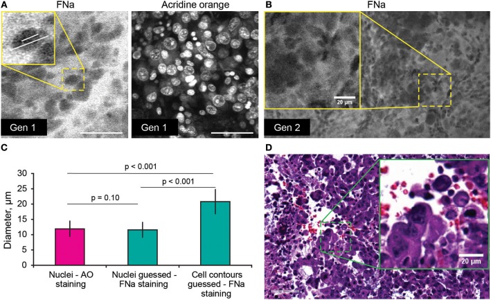
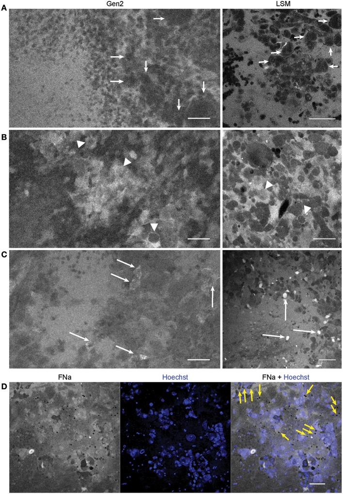
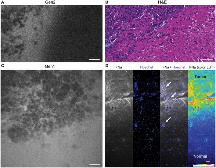
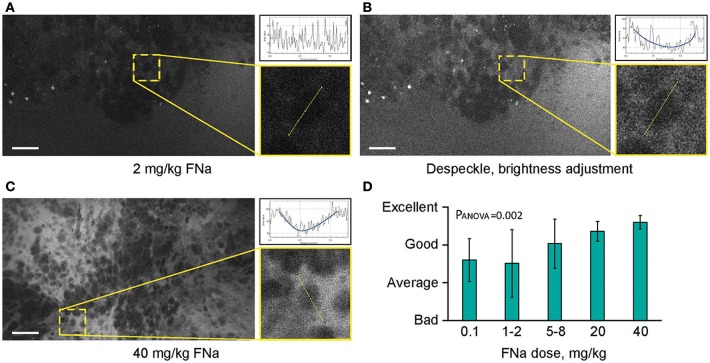
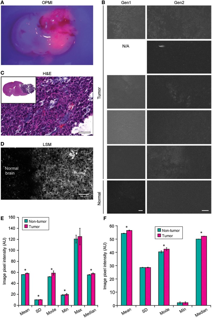
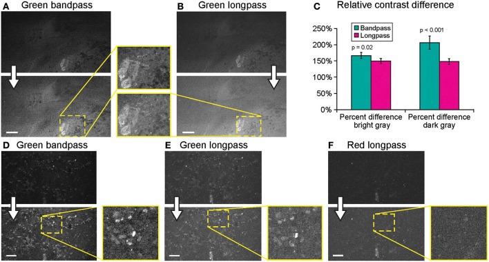
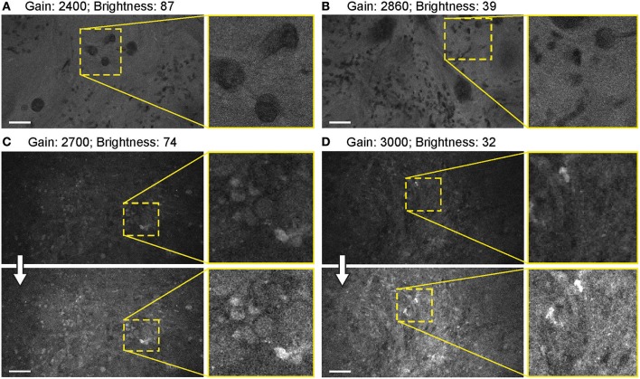
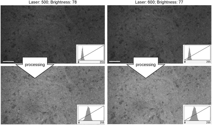
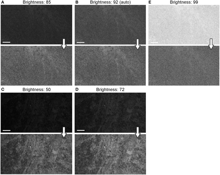
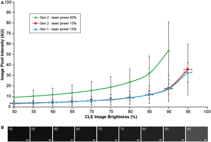
Previous studies showed that confocal laser endomicroscopy (CLE) images of brain tumors acquired by a first-generation (Gen1) CLE system using fluorescein sodium (FNa) contrast yielded a diagnostic accuracy similar to frozen surgical sections and histologic analysis. We investigated performance improvements of a second-generation (Gen2) CLE system designed specifically for neurosurgical use. Rodent glioma models were used for and rapid CLE imaging. FNa and 5-aminolevulinic acid were used as contrast agents. Gen1 and Gen2 CLE images were compared to distinguish cytoarchitectural features of tumor mass and margin and surrounding and normal brain regions. We assessed imaging parameters (gain, laser power, brightness, scanning speed, imaging depth, and Z-stack [3D image acquisition]) and evaluated optimal values for better neurosurgical imaging performance with Gen2. Efficacy of Gen1 and Gen2 was similar in identifying normal brain tissue, vasculature, and tumor cells in masses or at margins. Gen2 had smaller field of view, but higher image resolution, and sharper, clearer images. Other advantages of the Gen2 were auto-brightness correction, user interface, image metadata handling, and image transfer. CLE imaging with FNa allowed identification of nuclear and cytoplasmic contours in tumor cells. Injection of higher dosages of FNa (20 and 40 mg/kg vs. 0.1-8 mg/kg) resulted in better image clarity and structural identification. When used with 5-aminolevulinic acid, CLE was not able to detect individual glioma cells labeled with protoporphyrin IX, but overall fluorescence intensity was higher ( < 0.01) than in the normal hemisphere. Gen2 Z-stack imaging allowed a unique 3D image volume presentation through the focal depth. Compared with Gen1, advantages of Gen2 CLE included a more responsive and intuitive user interface, collection of metadata with each image, automatic Z-stack imaging, sharper images, and a sterile sheath. Shortcomings of Gen2 were a slightly slower maximal imaging speed and smaller field of view. Optimal Gen2 imaging parameters to visualize brain tumor cytoarchitecture with FNa as a fluorescent contrast were defined to aid further neurosurgical clinical and rapid use. Further validation of the Gen2 CLE for microscopic visualization and diagnosis of brain tumors is ongoing.
先前的研究表明,第一代(Gen1)共聚焦激光内镜(CLE)系统使用荧光素钠(FNa)造影剂获取的脑肿瘤CLE图像,其诊断准确性与冷冻手术切片及组织学分析相似。我们研究了专门为神经外科手术设计的第二代(Gen2)CLE系统的性能提升情况。使用啮齿动物胶质瘤模型进行快速CLE成像。FNa和5-氨基酮戊酸用作造影剂。比较Gen1和Gen2的CLE图像,以区分肿瘤肿块及边缘以及周围和正常脑区的细胞结构特征。我们评估了成像参数(增益、激光功率、亮度、扫描速度、成像深度和Z轴堆叠[三维图像采集]),并评估了Gen2实现更好神经外科成像性能的最佳值。Gen1和Gen2在识别正常脑组织、脉管系统以及肿块或边缘的肿瘤细胞方面效果相似。Gen2的视野较小,但图像分辨率更高,图像更清晰、锐利。Gen2的其他优点包括自动亮度校正、用户界面、图像元数据处理和图像传输。使用FNa进行CLE成像能够识别肿瘤细胞中的细胞核和细胞质轮廓。注射更高剂量的FNa(20和40mg/kg,对比0.1 - 8mg/kg)可使图像清晰度和结构识别效果更好。当与5-氨基酮戊酸一起使用时,CLE无法检测到用原卟啉IX标记的单个胶质瘤细胞,但总体荧光强度高于正常半球(P < 0.01)。Gen2的Z轴堆叠成像可通过焦深实现独特的三维图像容积呈现。与Gen1相比,Gen2 CLE的优点包括更灵敏、直观的用户界面、每张图像收集元数据、自动Z轴堆叠成像、图像更清晰以及无菌鞘。Gen2的缺点是最大成像速度稍慢且视野较小。确定了以FNa作为荧光造影剂可视化脑肿瘤细胞结构的Gen2最佳成像参数,以帮助进一步用于神经外科临床和快速应用。Gen2 CLE用于脑肿瘤微观可视化和诊断的进一步验证正在进行中。