• 文献检索
  • 文档翻译
  • 深度研究
  • 学术资讯
  • Suppr Zotero 插件Zotero 插件
  • 邀请有礼
  • 套餐&价格
  • 历史记录
应用&插件
Suppr Zotero 插件Zotero 插件浏览器插件Mac 客户端Windows 客户端微信小程序
定价
高级版会员购买积分包购买API积分包
服务
文献检索文档翻译深度研究API 文档MCP 服务
关于我们
关于 Suppr公司介绍联系我们用户协议隐私条款
关注我们

Suppr 超能文献

核心技术专利:CN118964589B侵权必究
粤ICP备2023148730 号-1Suppr @ 2026

文献检索

告别复杂PubMed语法,用中文像聊天一样搜索,搜遍4000万医学文献。AI智能推荐,让科研检索更轻松。

立即免费搜索

文件翻译

保留排版,准确专业,支持PDF/Word/PPT等文件格式,支持 12+语言互译。

免费翻译文档

深度研究

AI帮你快速写综述,25分钟生成高质量综述,智能提取关键信息,辅助科研写作。

立即免费体验

利用5-氨基乙酰丙酸诱导的原卟啉IX的高分辨率显微镜实现定量神经外科手术引导

Toward Quantitative Neurosurgical Guidance With High-Resolution Microscopy of 5-Aminolevulinic Acid-Induced Protoporphyrin IX.

作者信息

Wei Linpeng, Fujita Yoko, Sanai Nader, Liu Jonathan T C

机构信息

Department of Mechanical Engineering, University of Washington, Seattle, WA, United States.

Department of Neurological Surgery, Barrow Neurological Institute, Phoenix, AZ, United States.

出版信息

Front Oncol. 2019 Jul 3;9:592. doi: 10.3389/fonc.2019.00592. eCollection 2019.

DOI:10.3389/fonc.2019.00592
PMID:31334117
原文链接:https://pmc.ncbi.nlm.nih.gov/articles/PMC6616084/
Abstract

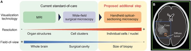
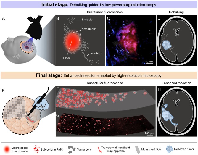
Low-power fluorescence microscopy of 5-ALA-induced PpIX has emerged as a valuable intraoperative imaging technology for improving the resection of malignant gliomas. However, current fluorescence imaging tools are not highly sensitive nor quantitative, which limits their effectiveness for optimizing operative decisions near the surgical margins of gliomas, in particular non-enhancing low-grade gliomas. Intraoperative high-resolution optical-sectioning microscopy can potentially serve as a valuable complement to low-power fluorescence microscopy by providing reproducible quantification of tumor parameters at the infiltrative margins of diffuse gliomas. In this forward-looking perspective article, we provide a brief discussion of recent technical advancements, pilot clinical studies, and our vision of the future adoption of handheld optical-sectioning microscopy at the final stages of glioma surgeries to enhance the extent of resection. We list a number of challenges for clinical acceptance, as well as potential strategies to overcome such obstacles for the surgical implementation of these microscopy techniques.

摘要

5-氨基乙酰丙酸(5-ALA)诱导的原卟啉IX(PpIX)的低倍荧光显微镜检查已成为一种有价值的术中成像技术,可用于改善恶性胶质瘤的切除效果。然而,目前的荧光成像工具既不高度敏感也不具备定量功能,这限制了它们在优化胶质瘤手术边缘(特别是无强化的低级别胶质瘤)手术决策方面的有效性。术中高分辨率光学切片显微镜检查通过对弥漫性胶质瘤浸润边缘的肿瘤参数进行可重复定量,有可能成为低倍荧光显微镜检查的重要补充。在这篇前瞻性观点文章中,我们简要讨论了近期的技术进展、初步临床研究,以及我们对在胶质瘤手术最后阶段采用手持式光学切片显微镜以提高切除范围的未来展望。我们列出了临床接受方面的一些挑战,以及克服这些障碍以实现这些显微镜技术手术应用的潜在策略。

https://cdn.ncbi.nlm.nih.gov/pmc/blobs/c267/6616084/c28cd6430c94/fonc-09-00592-g0002.jpg
https://cdn.ncbi.nlm.nih.gov/pmc/blobs/c267/6616084/c0d970e9df07/fonc-09-00592-g0001.jpg
https://cdn.ncbi.nlm.nih.gov/pmc/blobs/c267/6616084/c28cd6430c94/fonc-09-00592-g0002.jpg
https://cdn.ncbi.nlm.nih.gov/pmc/blobs/c267/6616084/c0d970e9df07/fonc-09-00592-g0001.jpg
https://cdn.ncbi.nlm.nih.gov/pmc/blobs/c267/6616084/c28cd6430c94/fonc-09-00592-g0002.jpg

相似文献

1
Toward Quantitative Neurosurgical Guidance With High-Resolution Microscopy of 5-Aminolevulinic Acid-Induced Protoporphyrin IX.利用5-氨基乙酰丙酸诱导的原卟啉IX的高分辨率显微镜实现定量神经外科手术引导
Front Oncol. 2019 Jul 3;9:592. doi: 10.3389/fonc.2019.00592. eCollection 2019.
2
Visualization technologies for 5-ALA-based fluorescence-guided surgeries.基于 5-ALA 的荧光引导手术的可视化技术。
J Neurooncol. 2019 Feb;141(3):495-505. doi: 10.1007/s11060-018-03077-9. Epub 2018 Dec 15.
3
Optical-sectioning microscopy of protoporphyrin IX fluorescence in human gliomas: standardization and quantitative comparison with histology.人胶质瘤原卟啉 IX 荧光的光学切片显微镜检查:与组织学的标准化和定量比较。
J Biomed Opt. 2017 Apr 1;22(4):46005. doi: 10.1117/1.JBO.22.4.046005.
4
Quantitative fluorescence using 5-aminolevulinic acid-induced protoporphyrin IX biomarker as a surgical adjunct in low-grade glioma surgery.使用5-氨基酮戊酸诱导的原卟啉IX生物标志物进行定量荧光作为低级别胶质瘤手术的手术辅助手段。
J Neurosurg. 2015 Sep;123(3):771-80. doi: 10.3171/2014.12.JNS14391. Epub 2015 Jul 3.
5
Dual-labeling with 5-aminolevulinic acid and fluorescein for fluorescence-guided resection of high-grade gliomas: technical note.5-氨基酮戊酸和荧光素双重标记在高级别胶质瘤荧光引导切除术中的应用:技术说明。
J Neurosurg. 2018 Feb;128(2):399-405. doi: 10.3171/2016.11.JNS161072. Epub 2017 Mar 24.
6
Comparing high-resolution microscopy techniques for potential intraoperative use in guiding low-grade glioma resections.比较高分辨率显微镜技术在低级别胶质瘤切除术中潜在的术中应用。
Lasers Surg Med. 2015 Apr;47(4):289-95. doi: 10.1002/lsm.22347. Epub 2015 Apr 14.
7
5-ALA in Suspected Low-Grade Gliomas: Current Role, Limitations, and New Approaches.5-氨基乙酰丙酸在疑似低级别胶质瘤中的应用:当前作用、局限性及新方法
Front Oncol. 2021 Jul 30;11:699301. doi: 10.3389/fonc.2021.699301. eCollection 2021.
8
5-aminolevulinic acid, fluorescein sodium, and indocyanine green for glioma margin detection: analysis of operating wide-field and confocal microscopy in glioma models of various grades.用于神经胶质瘤边缘检测的5-氨基乙酰丙酸、荧光素钠和吲哚菁绿:不同级别神经胶质瘤模型中的手术广角显微镜和共聚焦显微镜分析
Front Oncol. 2023 May 23;13:1156812. doi: 10.3389/fonc.2023.1156812. eCollection 2023.
9
Cadherin 13 overexpression as an important factor related to the absence of tumor fluorescence in 5-aminolevulinic acid-guided resection of glioma.钙黏蛋白 13 过表达是导致胶质瘤 5-氨基酮戊酸引导切除术中肿瘤荧光缺失的重要因素。
J Neurosurg. 2013 Nov;119(5):1331-9. doi: 10.3171/2013.7.JNS122340. Epub 2013 Sep 6.
10
Analysis of 5-aminolevulinic acid-induced fluorescence in 55 different spinal tumors.55 种不同脊髓肿瘤中 5-氨基酮戊酸诱导荧光的分析。
Neurosurg Focus. 2014 Feb;36(2):E11. doi: 10.3171/2013.12.FOCUS13485.

引用本文的文献

1
5-ALA localises to the autophagy compartment and increases its fluorescence upon autophagy enhancement through caloric restriction and spermidine treatment in human glioblastoma.5-氨基乙酰丙酸定位于自噬区室,并在人胶质母细胞瘤中通过热量限制和亚精胺处理增强自噬时增加其荧光。
Biochem Biophys Rep. 2024 Jan 13;37:101642. doi: 10.1016/j.bbrep.2024.101642. eCollection 2024 Mar.
2
The diagnostic accuracy of intraoperative differentiation and delineation techniques in brain tumours.脑肿瘤术中鉴别与勾勒技术的诊断准确性
Discov Oncol. 2022 Nov 10;13(1):123. doi: 10.1007/s12672-022-00585-z.
3
Turning on the light for brain tumor surgery: A 5-aminolevulinic acid story.

本文引用的文献

1
Dual-axis confocal microscopy for point-of-care pathology.用于即时护理病理学的双轴共聚焦显微镜。
IEEE J Sel Top Quantum Electron. 2019 Jan-Feb;25(1). doi: 10.1109/JSTQE.2018.2854572. Epub 2018 Jul 25.
2
Handheld line-scanned dual-axis confocal microscope with pistoned MEMS actuation for flat-field fluorescence imaging.手持线扫描双轴共焦显微镜,采用活塞式微机电系统(MEMS)驱动,用于平面荧光成像。
Opt Lett. 2019 Feb 1;44(3):671-674. doi: 10.1364/OL.44.000671.
3
Visualization technologies for 5-ALA-based fluorescence-guided surgeries.
脑肿瘤手术中的“明灯”:5-氨基酮戊酸的故事。
Neuro Oncol. 2022 Nov 2;24(Suppl 6):S52-S61. doi: 10.1093/neuonc/noac191.
4
In vivo microscopy as an adjunctive tool to guide detection, diagnosis, and treatment.体内显微镜作为一种辅助工具,用于指导检测、诊断和治疗。
J Biomed Opt. 2022 Apr;27(4). doi: 10.1117/1.JBO.27.4.040601.
5
Preliminary Report: Rapid Intraoperative Detection of Residual Glioma Cell in Resection Cavity Walls Using a Compact Fluorescence Microscope.初步报告:使用紧凑型荧光显微镜在切除腔壁中快速术中检测残留胶质瘤细胞
J Clin Med. 2021 Nov 18;10(22):5375. doi: 10.3390/jcm10225375.
6
Fluorescence Lifetime Imaging and Spectroscopic Co-Validation for Protoporphyrin IX-Guided Tumor Visualization in Neurosurgery.荧光寿命成像与光谱联合验证用于神经外科手术中原卟啉IX引导的肿瘤可视化
Front Oncol. 2021 Sep 14;11:741303. doi: 10.3389/fonc.2021.741303. eCollection 2021.
7
Combined autofluorescence and diffuse reflectance for brain tumour surgical guidance: initial study results.联合自体荧光和漫反射用于脑肿瘤手术导航:初步研究结果
Biomed Opt Express. 2021 Mar 29;12(4):2432-2446. doi: 10.1364/BOE.420292. eCollection 2021 Apr 1.
8
Harnessing non-destructive 3D pathology.利用非破坏性三维病理学
Nat Biomed Eng. 2021 Mar;5(3):203-218. doi: 10.1038/s41551-020-00681-x. Epub 2021 Feb 15.
9
5-aminolevulinic acid-guided surgery for focal pediatric brainstem gliomas: A preliminary study.5-氨基酮戊酸引导下的小儿局灶性脑干胶质瘤手术:一项初步研究。
Surg Neurol Int. 2020 Oct 8;11:334. doi: 10.25259/SNI_246_2020. eCollection 2020.
10
Video-Mosaicked Handheld Dual-Axis Confocal Microscopy of Gliomas: An Feasibility Study in Humans.胶质瘤的视频拼接手持式双轴共聚焦显微镜检查:一项人体可行性研究
Front Oncol. 2020 Aug 27;10:1674. doi: 10.3389/fonc.2020.01674. eCollection 2020.
基于 5-ALA 的荧光引导手术的可视化技术。
J Neurooncol. 2019 Feb;141(3):495-505. doi: 10.1007/s11060-018-03077-9. Epub 2018 Dec 15.
4
Evidence for Improving Outcome Through Extent of Resection.通过扩大切除范围改善预后的证据。
Neurosurg Clin N Am. 2019 Jan;30(1):85-93. doi: 10.1016/j.nec.2018.08.005. Epub 2018 Nov 1.
5
Utilization of intraoperative confocal laser endomicroscopy in brain tumor surgery.术中共聚焦激光内镜检查在脑肿瘤手术中的应用。
J Neurosurg Sci. 2018 Dec;62(6):704-717. doi: 10.23736/S0390-5616.18.04553-8. Epub 2018 Aug 28.
6
Light-sheet microscopy for slide-free non-destructive pathology of large clinical specimens.用于大型临床标本的无玻片非破坏性病理学的光片显微镜技术。
Nat Biomed Eng. 2017 Jul;1(7). doi: 10.1038/s41551-017-0084. Epub 2017 Jun 26.
7
5-ALA in the management of malignant glioma.5-氨基乙酰丙酸在恶性胶质瘤治疗中的应用
Lasers Surg Med. 2018 Jul;50(5):399-419. doi: 10.1002/lsm.22933. Epub 2018 May 8.
8
Correlation Between the Residual Tumor Volume, Extent of Tumor Resection, and O-Methylguanine DNA Methyltransferase Status in Patients with Glioblastoma.胶质母细胞瘤患者残余肿瘤体积、肿瘤切除范围与O-甲基鸟嘌呤-DNA甲基转移酶状态之间的相关性
World Neurosurg. 2018 Aug;116:e147-e161. doi: 10.1016/j.wneu.2018.04.134. Epub 2018 Apr 27.
9
Spectral Radiance of Protoporphyrin IX Fluorescence and Its Histopathological Implications in 5-Aminolevulinic Acid-Guided Surgery for Glioblastoma.原卟啉IX荧光的光谱辐射及其在5-氨基酮戊酸引导的胶质母细胞瘤手术中的组织病理学意义
Photomed Laser Surg. 2018 May;36(5):266-272. doi: 10.1089/pho.2017.4384. Epub 2018 Feb 26.
10
Scanning Fiber Endoscope Improves Detection of 5-Aminolevulinic Acid-Induced Protoporphyrin IX Fluorescence at the Boundary of Infiltrative Glioma.扫描光纤内窥镜可提高对浸润性胶质瘤边界处5-氨基酮戊酸诱导的原卟啉IX荧光的检测。
World Neurosurg. 2018 May;113:e51-e69. doi: 10.1016/j.wneu.2018.01.151. Epub 2018 Feb 2.