Department of Biomedical and Molecular Science, Queen's University, Kingston, Ontario, Canada.
PLoS One. 2019 Jul 23;14(7):e0220045. doi: 10.1371/journal.pone.0220045. eCollection 2019.
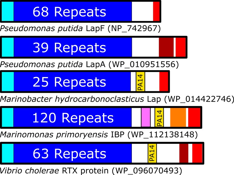
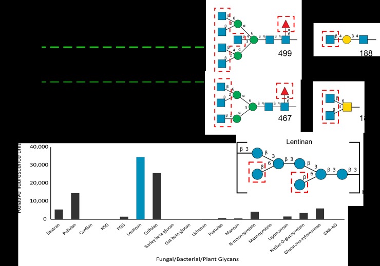
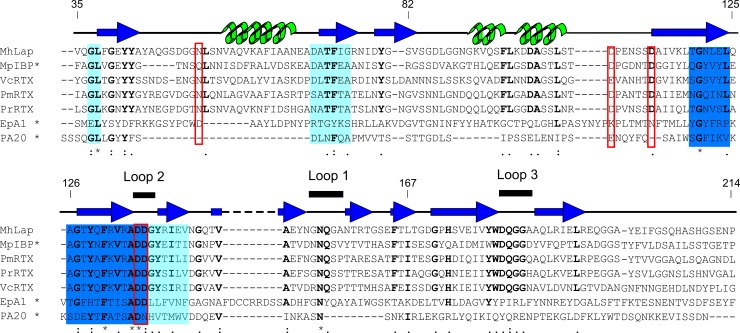
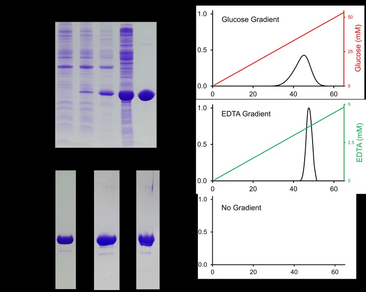
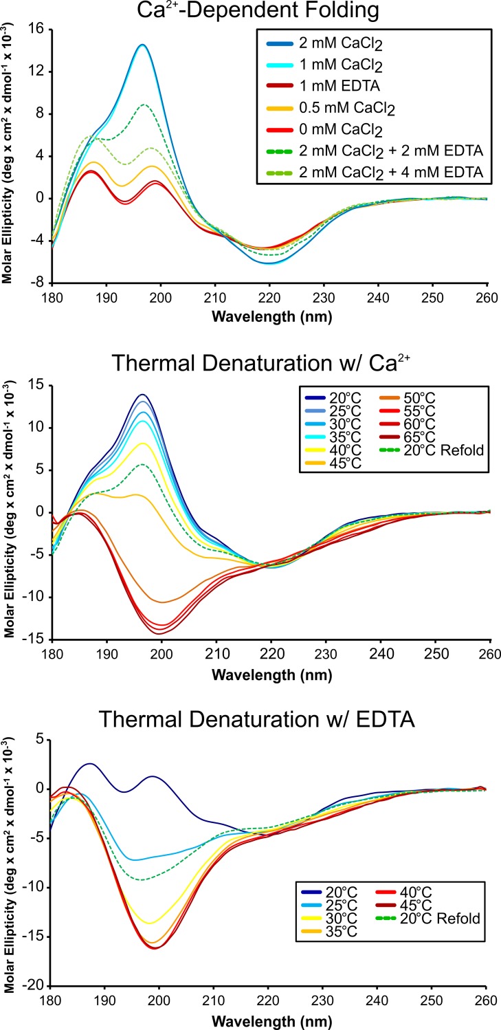
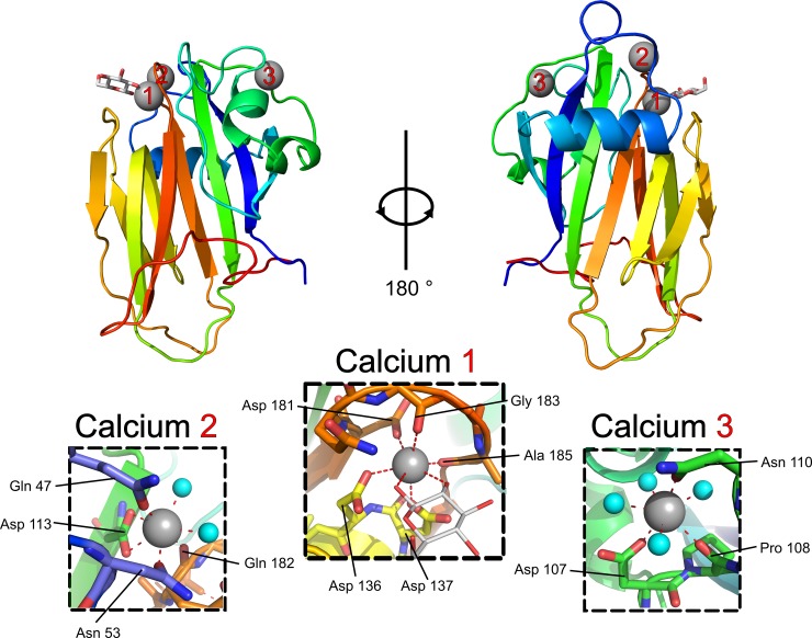
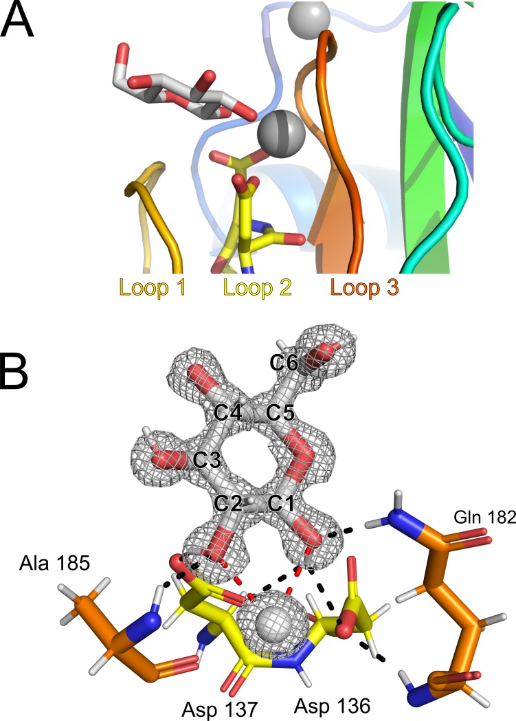
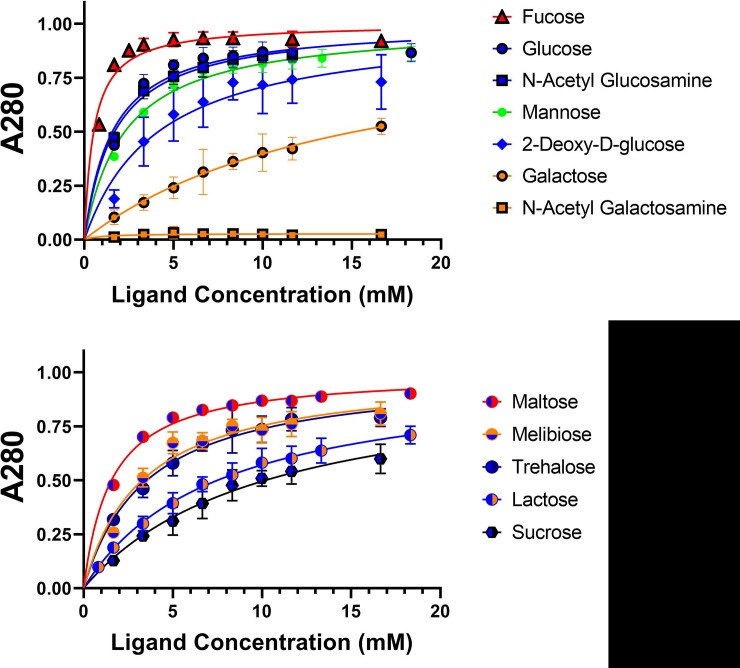
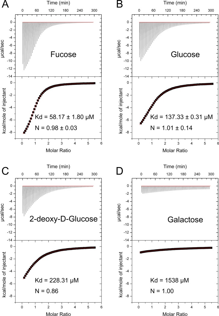
Bacterial adhesins attach their hosts to surfaces through one or more ligand-binding domains. In RTX adhesins, which are localized to the outer membrane of many Gram-negative bacteria via the type I secretion system, we see several examples of a putative sugar-binding domain. Here we have recombinantly expressed one such ~20-kDa domain from the ~340-kDa adhesin found in Marinobacter hydrocarbonoclasticus, an oil-degrading bacterium. The sugar-binding domain was purified from E. coli with a yield of 100 mg/L of culture. Circular dichroism analysis showed that the protein was rich in beta-structure, was moderately heat resistant, and required Ca2+ for proper folding. A crystal structure was obtained in Ca2+ at 1.2-Å resolution, which showed the presence of three Ca2+ ions, two of which were needed for structural integrity and one for binding sugars. Glucose was soaked into the crystal, where it bound to the sugar's two vicinal hydroxyl groups attached to the first and second (C1 and C2) carbons in the pyranose ring. This attraction to glucose caused the protein to bind certain polysaccharide-based column matrices and was used in a simple competitive binding assay to assess the relative affinity of sugars for the protein's ligand-binding site. Fucose, glucose and N-acetylglucosamine bound most tightly, and N-acetylgalactosamine hardly bound at all. Isothermal titration calorimetry was used to determine specific binding affinities, which lie in the 100-μM range. Glycan arrays were tested to expand the range of ligand sugars assayed, and showed that MhPA14 bound preferentially to branched polymers containing terminal sugars highlighted as strong binders in the competitive binding assay. Some of these binders have vicinal hydroxyl groups attached to the C3 and C4 carbons that are sterically equivalent to those presented by the C1 and C2 carbons of glucose.
细菌黏附素通过一个或多个配体结合结构域将宿主附着到表面上。在 RTX 黏附素中,我们看到了几个假定的糖结合结构域的例子,这些黏附素通过 I 型分泌系统定位于许多革兰氏阴性菌的外膜上。在这里,我们从油降解菌 Marinobacter hydrocarbonoclasticus 中发现的340 kDa 黏附素中重组表达了一个这样的20 kDa 结构域。该糖结合结构域从大肠杆菌中以 100mg/L 培养物的产量得到纯化。圆二色性分析表明,该蛋白富含β-结构,具有中等耐热性,并且需要 Ca2+ 才能正确折叠。在 Ca2+ 存在下获得了 1.2-Å 分辨率的晶体结构,表明存在三个 Ca2+ 离子,其中两个对于结构完整性,一个对于结合糖是必需的。将葡萄糖浸泡到晶体中,在那里它与吡喃糖环的第一个和第二个(C1 和 C2)碳原子上的两个相邻羟基结合。这种对葡萄糖的吸引力导致该蛋白结合某些基于多糖的柱基质,并用于简单的竞争性结合测定法中,以评估糖对蛋白配体结合位点的相对亲和力。岩藻糖、葡萄糖和 N-乙酰葡萄糖胺结合得最紧密,而 N-乙酰半乳糖胺几乎完全不结合。等温滴定量热法用于确定特定的结合亲和力,其范围在 100 μM 左右。糖芯片用于扩展测定的配体糖的范围,并表明 MhPA14 优先结合含有末端糖的支链聚合物,这些末端糖在竞争性结合测定中被鉴定为强结合物。这些结合物中的一些具有与 C3 和 C4 碳原子连接的相邻羟基,这些碳原子在空间上与葡萄糖的 C1 和 C2 碳原子等效。