Biomarkers Unit, Department of Applied Research and Technical Development, Fondazione IRCCS Istituto Nazionale dei Tumori, Milan, Italy.
Radiation Oncology 1, Fondazione IRCCS Istituto Nazionale dei Tumori, Milan, Italy.
J Exp Clin Cancer Res. 2019 Jul 23;38(1):326. doi: 10.1186/s13046-019-1325-6.
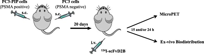
Prostate cancer (PCa) is the second leading cause of cancer-related death in the Western population. The use in oncology of positron emission tomography/computed tomography (PET/CT) with emerging radiopharmaceuticals promises accurate staging of primary disease, restaging of recurrent disease and detection of metastatic lesions. Prostate-specific membrane antigen (PSMA) expression, directly related to androgen-independence, metastasis and progression, renders this tumour associate antigen a good target for the development of new radiopharmaceuticals for PET. Aim of this study was to demonstrate in a preclinical in vivo model (PSMA-positive versus PSMA-negative tumours) the targeting specificity and sensitivity of the anti-PSMA single-chain variable fragment (scFv) labelled with I.
The I-labeling conditions of the antibody fragment scFvD2B were optimized and assessed for purity and immunoreactivity. The specificity of I-scFvD2B was tested in mice bearing PSMA-positive and PSMA-negative tumours to assess both ex-vivo biodistribution and immune-PET.
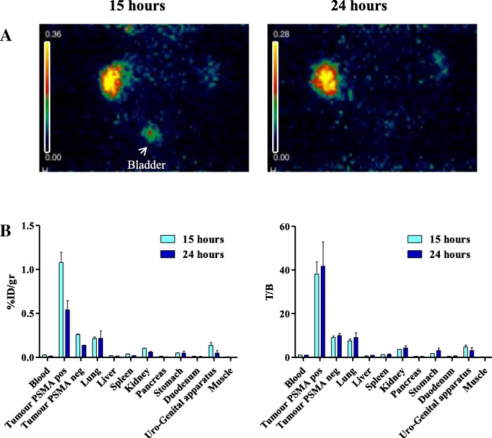
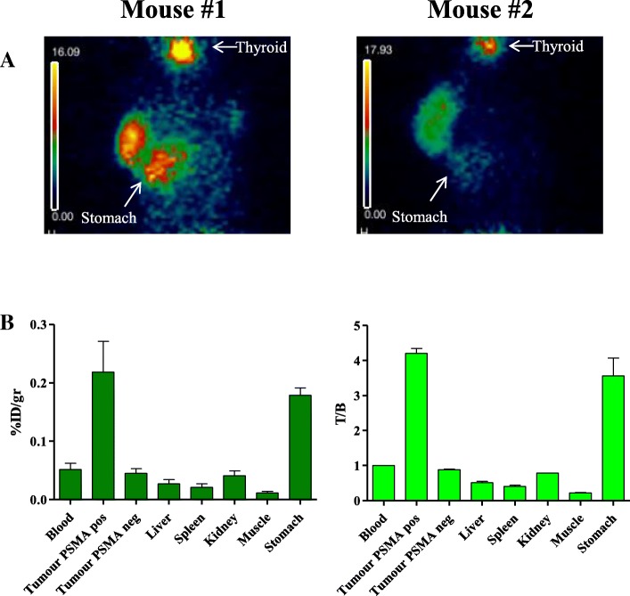
The uptake fraction of I-scFvD2B was very high on PSMA positive cells (range 75-91%) and highly specific and immuno-PET at the optimal time point, defined between 15 h and 24 h, provides a specific localization of lesions bearing the target antigen of interest (PSMA positive vs PSMA negative tumors %ID/g: p = 0.0198 and p = 0.0176 respectively) yielding a median target/background ratio around 30-40.
Preclinical in vivo results of our immuno-PET reagent are highly promising. The target to background ratio is improved notably using PET compared to SPECT previously performed. These data suggest that, upon clinical confirmation of sensitivity and specificity, our anti-PSMA I-scFvD2B may be superior to other diagnostic modalities for PCa. The possibility to combine in patients our I-scFvD2B in multi-modal systems, such as PET/CT, PET/MR and PET/SPECT/CT, will provide quantitative 3D tomographic images improving the knowledge of cancer biology and treatment.
前列腺癌(PCa)是西方人群癌症相关死亡的第二大原因。新兴放射性药物正电子发射断层扫描/计算机断层扫描(PET/CT)在肿瘤学中的应用有望准确分期原发性疾病、复发性疾病的分期和转移病灶的检测。前列腺特异性膜抗原(PSMA)的表达与雄激素独立性、转移和进展直接相关,使这种肿瘤相关抗原成为开发用于 PET 的新型放射性药物的良好靶标。本研究的目的是在临床前体内模型(PSMA 阳性与 PSMA 阴性肿瘤)中证明抗 PSMA 单链可变片段(scFv)标记的 I 的靶向特异性和敏感性。
优化了抗体片段 scFvD2B 的 I 标记条件,并评估了其纯度和免疫反应性。在携带 PSMA 阳性和 PSMA 阴性肿瘤的小鼠中测试了 I-scFvD2B 的特异性,以评估体外生物分布和免疫 PET。
I-scFvD2B 在 PSMA 阳性细胞上的摄取分数非常高(范围为 75-91%),且高度特异,在最佳时间点(定义为 15 小时至 24 小时之间)进行免疫 PET 可特异性定位携带目标抗原的病变(PSMA 阳性与 PSMA 阴性肿瘤 %ID/g:p=0.0198 和 p=0.0176),产生约 30-40 的中位靶标/背景比。
我们的免疫 PET 试剂的临床前体内结果非常有希望。与之前进行的 SPECT 相比,使用 PET 显著提高了靶标与背景的比值。这些数据表明,在临床确认敏感性和特异性后,我们的抗 PSMA I-scFvD2B 可能优于其他用于 PCa 的诊断方式。在患者中,我们的 I-scFvD2B 可以与多模态系统(如 PET/CT、PET/MR 和 PET/SPECT/CT)相结合,提供定量的 3D 断层图像,从而提高对癌症生物学和治疗的认识。