Han Hyun-Jung, Kim Jung-Hyun
Department of Veterinary Emergency Medicine, Konkuk Veterinary Medical Teaching Hospital, Konkuk University, Seoul, 05029, Republic of Korea.
Department of Veterinary Internal Medicine, College of Veterinary Medicine, Konkuk University, Seoul, 05029, Republic of Korea.
Acta Vet Scand. 2019 Jul 26;61(1):37. doi: 10.1186/s13028-019-0472-2.
Pulmonary hypoplasia (PH) and congenital lobar emphysema (CLE) are very rare congenital pulmonary anomalies in veterinary medicine. PH refers to the incomplete pulmonary development due to embryologic imbalance of bronchial development between the lung buds, while CLE is defined as alveolar hyperinflation due to bronchial collapse during expiration caused by bronchial cartilage dysplasia, external bronchial compression, and idiopathic etiology. CLE may develop into pulmonary blebs or bullae that may rupture and induce a spontaneous pneumothorax. There are no reports on concurrent PH and CLE in animals.
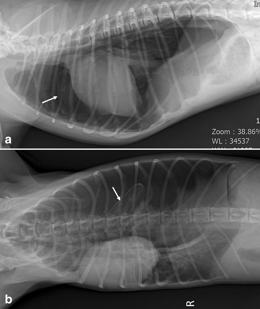
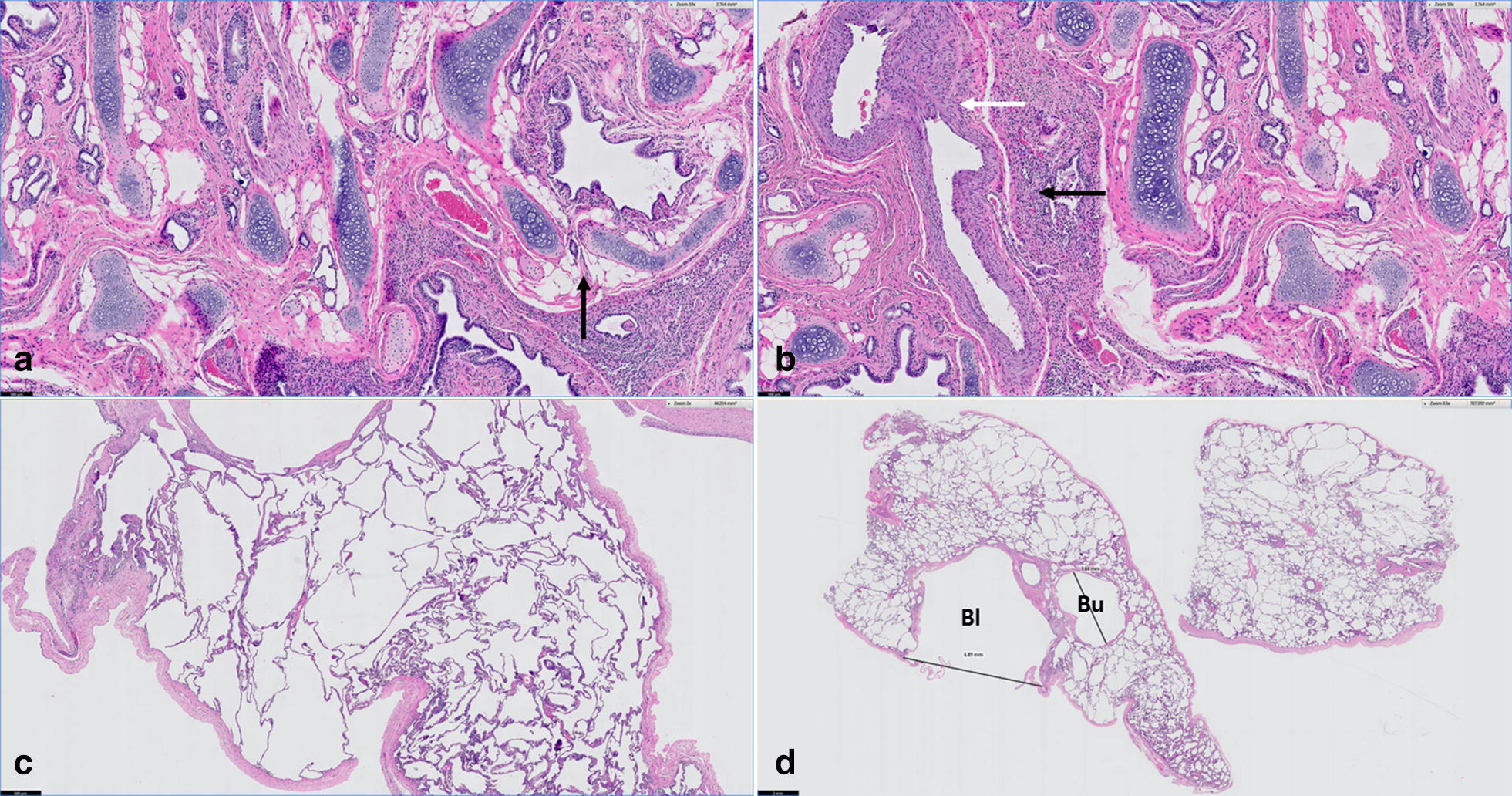
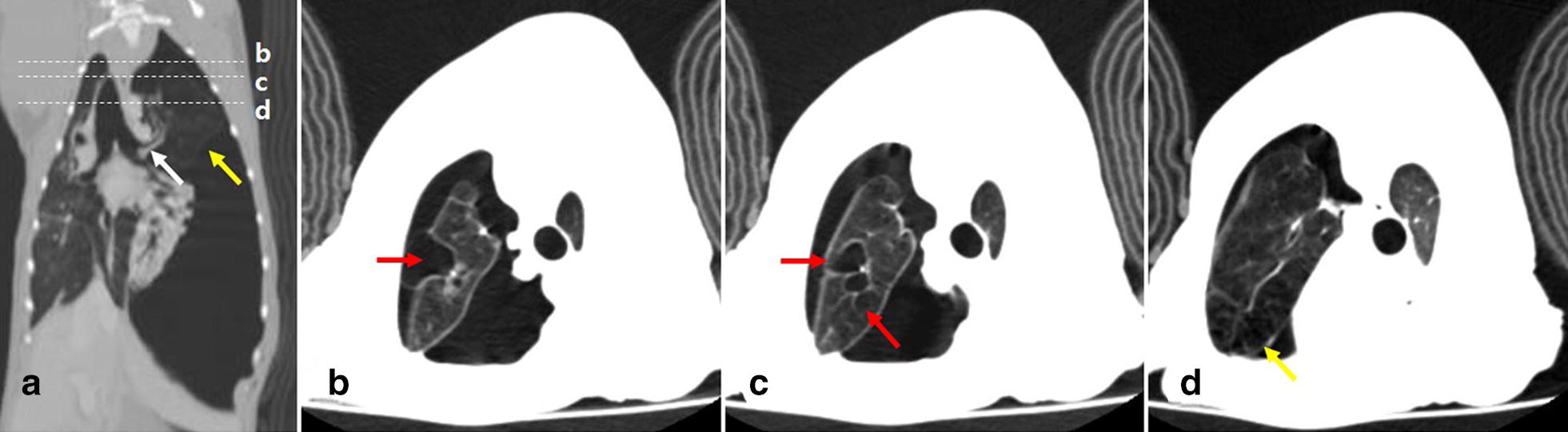
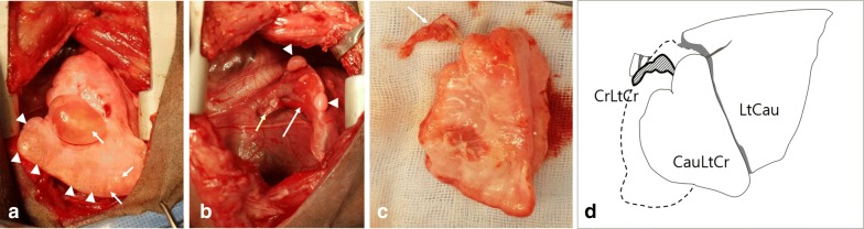
A 7-month-old castrated male Italian Greyhound weighing 5.5 kg presented with vomiting and acute onset of severe dyspnea without any previous history of disease. After emergency treatment including oxygen supplementation and thoracocentesis, plain radiology and computed tomography scanning were performed and lobar emphysema with multiple bullae in the left cranial lung lobe associated with tension pneumothorax was identified. Since the pneumothorax was not resolved despite continuous suction of intrathoracic air for 3 days, a complete lobectomy of the left cranial lung lobe was performed. The excised lobe was not grossly divided into cranial and caudal parts, but a tissue mass less than 1 cm in size was present at the hilum and cranial to the excised lobe. Postoperatively, the dog recovered rapidly without air retention in the thoracic cavity. Histopathologically, the mass was identified as a hypoplastic lung tissue with collapsed alveoli, bronchial dysplasia, and pulmonary arterial hypertrophy. Additionally, the excised lung lobe presented CLE with marked ectasia of alveoli, various blebs and bullae, and general bronchial cartilage dysplasia. According to gross and histopathologic findings, the dog was diagnosed with concurrent PH and CLE in the left cranial lung lobe. During 16 months of follow-up, the dog was well and without any respiratory problems.
This case report confirmed the clinical and histologic features of two different types of rare congenital pulmonary anomalies, PH and CLE, which occurred concurrently in a single lung lobe of a young dog. The condition was successfully managed with lobectomy.
肺发育不全(PH)和先天性肺叶气肿(CLE)在兽医学中是非常罕见的先天性肺部异常。PH是指由于肺芽之间支气管发育的胚胎学失衡导致的肺发育不完全,而CLE的定义是由于支气管软骨发育异常、外部支气管压迫和特发性病因导致呼气时支气管塌陷引起的肺泡过度充气。CLE可能发展为肺大疱或肺气泡,可能破裂并导致自发性气胸。目前尚无动物同时发生PH和CLE的报道。
一只7个月大、体重5.5千克的去势雄性意大利灵缇犬,此前无任何疾病史,出现呕吐和急性严重呼吸困难。在进行包括吸氧和胸腔穿刺在内的紧急治疗后,进行了X线平片和计算机断层扫描,发现左前肺叶有肺叶气肿并伴有多个肺大疱,同时伴有张力性气胸。尽管连续3天持续抽吸胸腔内气体,但气胸仍未缓解,遂对左前肺叶进行了完整的肺叶切除术。切除的肺叶在大体上未明显分为头侧和尾侧部分,但在肺门处及切除肺叶的头侧有一个大小小于1厘米的组织块。术后,该犬恢复迅速,胸腔内无气体潴留。组织病理学检查显示,该组织块为发育不全的肺组织,肺泡塌陷、支气管发育异常和肺动脉肥大。此外,切除的肺叶呈现CLE,肺泡明显扩张,有各种肺大疱和肺气泡,以及普遍的支气管软骨发育异常。根据大体和组织病理学检查结果,该犬被诊断为左前肺叶同时发生PH和CLE。在16个月的随访期间,该犬健康状况良好,无任何呼吸问题。
本病例报告证实了两种不同类型的罕见先天性肺部异常,即PH和CLE,在一只幼犬的单个肺叶中同时发生的临床和组织学特征。通过肺叶切除术成功治疗了该病症。