McKavanagh Rebecca, Torso Mario, Jenkinson Mark, Kolasinski James, Stagg Charlotte J, Esiri Margaret M, McNab Jennifer A, Johansen-Berg Heidi, Miller Karla L, Chance Steven A
Nuffield Department of Clinical Neurosciences, University of Oxford, Oxford, United Kingdom.
Wellcome Centre for Integrative Neuroimaging, FMRIB, Nuffield Department of Clinical Neurosciences, University of Oxford, Oxford, United Kingdom.
Hum Brain Mapp. 2019 Oct 15;40(15):4417-4431. doi: 10.1002/hbm.24711. Epub 2019 Jul 29.
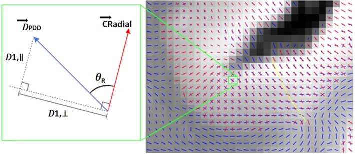
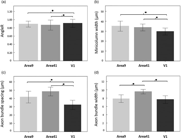
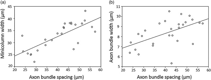
To investigate whether the observed anisotropic diffusion in cerebral cortex may reflect its columnar cytoarchitecture and myeloarchitecture, as a potential biomarker for disease-related changes, we compared postmortem diffusion magnetic resonance imaging scans of nine multiple sclerosis brains with histology measures from the same regions. Histology measurements assessed the cortical minicolumnar structure based on cell bodies and associated axon bundles in dorsolateral prefrontal cortex (Area 9), Heschl's gyrus (Area 41), and primary visual cortex (V1). Diffusivity measures included mean diffusivity, fractional anisotropy of the cortex, and three specific measures that may relate to the radial minicolumn structure: the angle of the principal diffusion direction in the cortex, the component that was perpendicular to the radial direction, and the component that was parallel to the radial direction. The cellular minicolumn microcircuit features were correlated with diffusion angle in Areas 9 and 41, and the axon bundle features were correlated with angle in Area 9 and to the parallel component in V1 cortex. This may reflect the effect of minicolumn microcircuit organisation on diffusion in the cortex, due to the number of coherently arranged membranes and myelinated structures. Several of the cortical diffusion measures showed group differences between MS brains and control brains. Differences between brain regions were also found in histology and diffusivity measurements consistent with established regional variation in cytoarchitecture and myeloarchitecture. Therefore, these novel measures may provide a surrogate of cortical organisation as a potential biomarker, which is particularly relevant for detecting regional changes in neurological disorders.
为了研究在大脑皮层中观察到的各向异性扩散是否可能反映其柱状细胞结构和髓鞘结构,作为疾病相关变化的潜在生物标志物,我们将9例多发性硬化症患者大脑的死后扩散磁共振成像扫描结果与同一区域的组织学测量结果进行了比较。组织学测量基于背外侧前额叶皮层(9区)、颞横回(41区)和初级视觉皮层(V1)中的细胞体和相关轴突束评估皮质微柱结构。扩散率测量包括平均扩散率、皮层的分数各向异性,以及可能与径向微柱结构相关的三个特定测量值:皮层中主扩散方向的角度、垂直于径向的分量和平行于径向的分量。细胞微柱微电路特征与9区和41区的扩散角相关,轴突束特征与9区的角度和V1皮层中的平行分量相关。这可能反映了微柱微电路组织对皮层扩散的影响,这是由于相干排列的膜和髓鞘结构的数量所致。几种皮层扩散测量显示多发性硬化症患者大脑与对照大脑之间存在组间差异。在组织学和扩散率测量中也发现了脑区之间的差异,这与细胞结构和髓鞘结构中既定的区域差异一致。因此,这些新的测量方法可能提供一种皮层组织的替代指标作为潜在的生物标志物,这对于检测神经系统疾病中的区域变化尤为重要。