Rajagopal Niranjana, Yamada Yasuhiro, Balaji Arun, Kawase Tsukasa, Kato Yoko
Department of Neurosurgery, Sri Sathya Sai Institute of Higher Medical Sciences, Bangalore, India.
Department of Neurosurgery, Fujita Health University, Banbuntane Hotokukai Hospital, Nagoya, Japan.
Int J Surg Case Rep. 2019;61:141-146. doi: 10.1016/j.ijscr.2019.07.037. Epub 2019 Jul 22.
Mirror aneurysms are a rare subtype of multiple aneurysms, located in identical or adjacent arterial segment bilaterally. We report a case series of 3 such patients amongst whom one of them had 3 sets of mirror aneurysms and the other patient had 2 sets of mirror aneurysm on the same arterial segment which has not been reported till date.
A retrospective analysis of 3 patients with incidentally detected multiple mirror aneurysms, who were treated with microsurgical clipping and coiling, was conducted. A systematic search was performed using the PUBMED database and relevant articles were reviewed with particular attention to incidence, associated conditions, risk factors and management strategies. Written informed consent was obtained from all of the patients for publication of this case report and accompanying images. A copy of the written consent is available for review by the Editor-in-Chief of this journal on request. This research work has been reported in line with the PROCESS criteria (Agha et al., 2018).
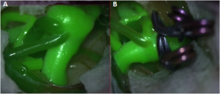
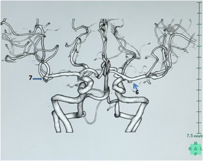
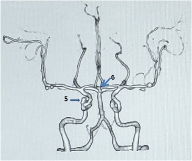
All the 3 patients were females with incidentally detected multiple mirror aneurysms. All the 3 patients were known Hypertensives. All the aneurysms were successfully clipped by a multistage, bilateral craniotomy except the one in the cavernous ICA, for which an endovascular procedure is planned. All of them had an uneventful postoperative course with the CT angiogram showing obliteration of all the clipped aneurysms except the one in the cavernous ICA.
Multiple mirror aneurysms represent a rare occurrence of a diverse pathology. Both these described types of cases have not been reported so far in the literature. The treatment strategy for mirror aneurysms should be determined individually based on the location, size, and morphology of the aneurysms, as well as, on the clinical manifestations of each patient.
镜像动脉瘤是多发性动脉瘤的一种罕见亚型,双侧位于相同或相邻动脉节段。我们报告了3例这样的患者,其中1例有3组镜像动脉瘤,另1例在同一动脉节段有2组镜像动脉瘤,这在迄今为止尚未见报道。
对3例偶然发现的多发性镜像动脉瘤患者进行回顾性分析,这些患者接受了显微手术夹闭和栓塞治疗。使用PubMed数据库进行系统检索,并对相关文章进行综述,特别关注发病率、相关情况、危险因素和治疗策略。获得了所有患者的书面知情同意,以发表本病例报告及附带图像。如有需要,本杂志主编可查阅书面同意书副本。本研究工作已按照PROCESS标准(Agha等人,2018年)进行报告。
所有3例患者均为女性,偶然发现多发性镜像动脉瘤。所有3例患者均为已知高血压患者。除海绵窦段颈内动脉的动脉瘤计划采用血管内治疗外,所有动脉瘤均通过多阶段双侧开颅手术成功夹闭。所有患者术后病程平稳,CT血管造影显示除海绵窦段颈内动脉的动脉瘤外,所有夹闭的动脉瘤均已闭塞。
多发性镜像动脉瘤是一种罕见的多种病理类型。这两种所述类型的病例在文献中迄今均未见报道。镜像动脉瘤的治疗策略应根据动脉瘤的位置、大小和形态以及每位患者的临床表现单独确定。