Dept. of Psychiatry and the Behavioral Sciences, Keck School of Medicine, University of Southern California, Los Angeles, CA, 90089, United States; Dept. of Neurology, Keck School of Medicine, University of Southern California, Los Angeles, CA, 90089, United States; Viterbi School of Engineering, Dept. of Biomedical Engineering, Los Angeles, CA, 90033, United States.
Viterbi School of Engineering, Dept. of Biomedical Engineering, Los Angeles, CA, 90033, United States.
Behav Brain Res. 2019 Dec 16;375:112116. doi: 10.1016/j.bbr.2019.112116. Epub 2019 Aug 1.
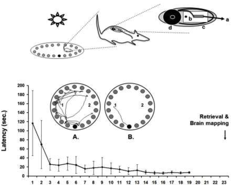
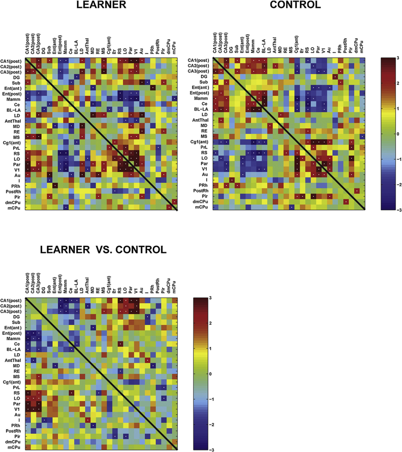
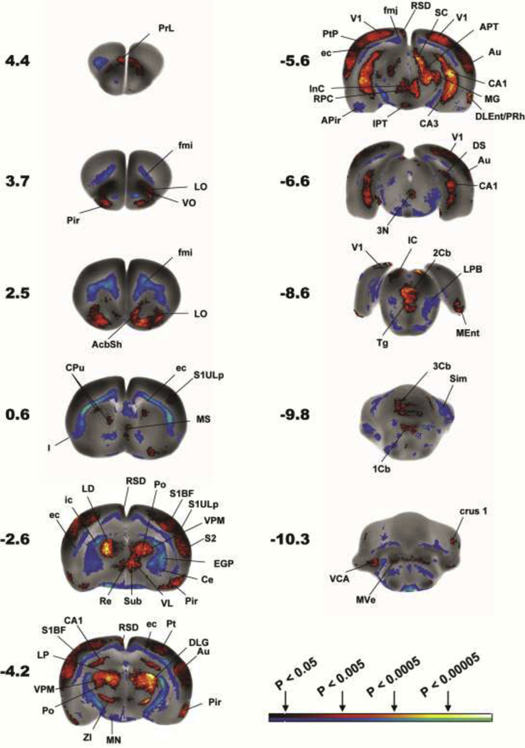
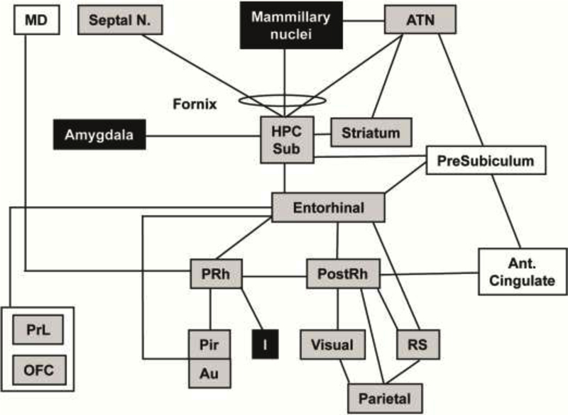
Studies of brain functional activation during spatial navigation using electrophysiology and immediate-early gene responses have typically targeted a limited number of brain regions. Our study provides the first whole brain analysis of cerebral activation during retrieval of spatial memory in the freely-moving rat. Rats (LEARNERS) were trained in the Barnes maze, an allocentric spatial navigation task, while CONTROLS received passive exposure. After 19 days, functional brain mapping was performed during recall by bolus intravenous injection of [C]-iodoantipyrine using a novel subcutaneous minipump triggered by remote activation. Regional cerebral blood flow (rCBF)-related tissue radioactivity was analyzed by statistical parametric mapping from autoradiographic images of the three-dimensionally reconstructed brains. Functional connectivity was examined between regions of the spatial navigation circuit through interregional correlation analysis. Significant rCBF increases were noted in LEARNERS compared to CONTROLS broadly across the spatial navigation circuit, including the hippocampus (anterior dorsal CA1, posterior ventral CA1-3), subiculum, thalamus, striatum, medial septum, cerebral cortex, with decreases noted in the mammillary nucleus, amygdala and insula. LEARNERS showed a significantly greater positive correlation of rCBF of the ventral hippocampus with retrosplenial, lateral orbital, parietal and primary visual cortex, and a significantly more negative correlation with the mammillary nucleus, amygdala, posterior entorhinal cortex, and anterior thalamic nucleus. The complex sensory component of the spatial navigation task was underscored by broad activation across visual, somatosensory, olfactory, auditory and vestibular circuits which was enhanced in LEARNERS. Brain mapping facilitated by an implantable minipump represents a powerful tool for evaluation of mammalian behaviors dependent on locomotion.
使用电生理学和即刻早期基因反应研究空间导航过程中的大脑功能激活,通常针对的是数量有限的大脑区域。我们的研究提供了在自由移动的大鼠中检索空间记忆过程中大脑激活的全脑分析。大鼠(学习者)在 Barnes 迷宫中接受训练,这是一种与位置有关的空间导航任务,而对照组则接受被动暴露。19 天后,通过使用新型皮下微泵进行的静脉内碘安替比林推注,在回忆过程中进行功能脑映射,该微泵由远程激活触发。通过对三维重建大脑的放射自显影图像进行统计参数映射,分析与区域脑血流(rCBF)相关的组织放射性。通过区域间相关分析,检查空间导航回路中各区域之间的功能连接。与对照组相比,学习者在空间导航回路的广泛区域中 rCBF 显著增加,包括海马体(前背 CA1、后背 CA1-3)、下托、丘脑、纹状体、内侧隔核、大脑皮层,而在乳头核、杏仁核和岛叶中 rCBF 减少。学习者的腹侧海马体 rCBF 与后隔叶、外侧眶额、顶叶和初级视觉皮层的正相关显著增加,与乳头核、杏仁核、后内嗅皮质和前丘脑核的负相关显著增加。空间导航任务的复杂感觉成分在视觉、躯体感觉、嗅觉、听觉和前庭回路中广泛激活,在学习者中得到增强。植入式微泵促进的脑映射代表了评估依赖于运动的哺乳动物行为的有力工具。