Center for the Neurobiology of Stress, Department of Medicine, University of California, Los Angeles, CA, USA Department of Psychiatry and the Behavioral Sciences, University of Southern California, Los Angeles, CA, USA Veterans Administration, Greater Los Angeles Healthcare System, Los Angeles, CA, USA Department of Chemistry and Biochemistry, California State University, Los Angeles, CA, USA Program in Neuroscience, University of Southern California, Los Angeles, CA, USA Department of Biomedical Engineering, University of Southern California, Los Angeles, CA, USA Departments of Physiology, Psychiatry and Biobehavioral Sciences, Brain Research Institute, University of California, Los Angeles, CA, USA Departments of Cell and Neurobiology, Neurology, University of Southern California, Los Angeles, CA, USA.
Pain. 2011 Dec;152(12):2746-2756. doi: 10.1016/j.pain.2011.08.022. Epub 2011 Sep 22.
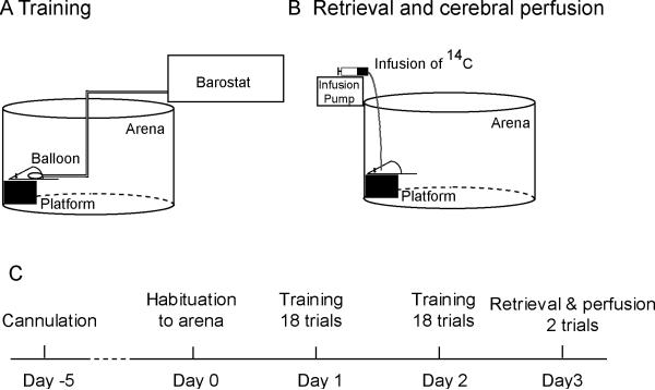
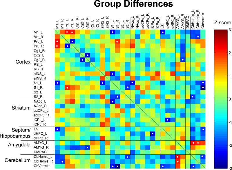
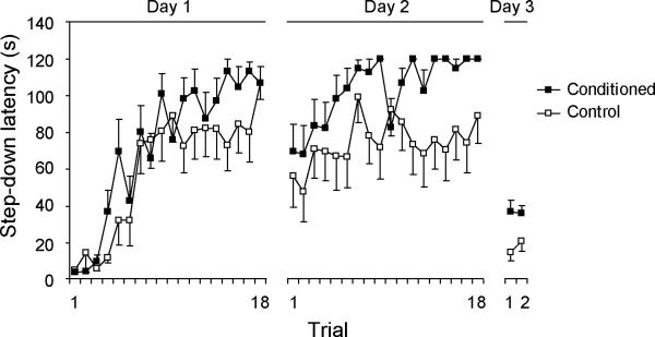
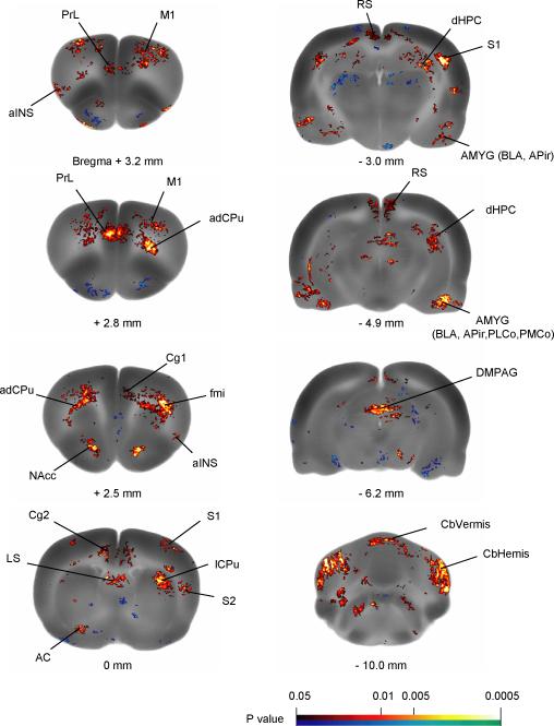
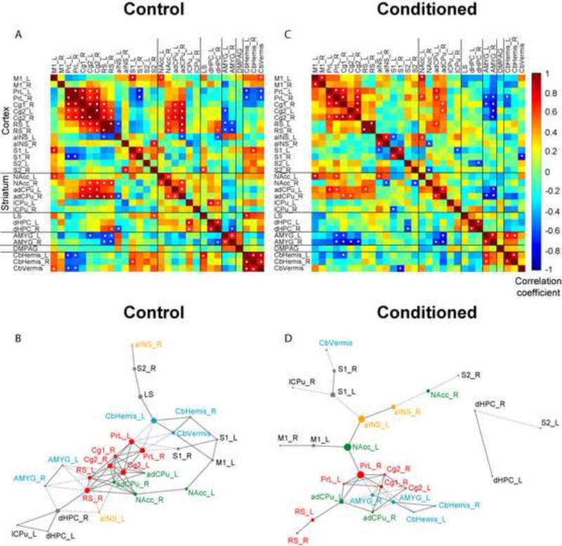
This study assessed functional brain activation in rats during expectation of visceral pain. Male rats were trained in step-down passive avoidance (PA) for 2 days. Upon stepping down from a platform, conditioned animals received noxious colorectal distension delivered through a colorectal balloon, whereas the balloon in control rats remained uninflated. On day 3, PA behavior was assessed while [(14)C]-iodoantipyrine was infused intravenously, followed by immediate euthanasia. Regional cerebral blood flow-related tissue radioactivity (rCBF) was analyzed by statistical parametric mapping using 3-dimensional brains reconstructed from autoradiographic brain slice images. Associated with retrieved PA behavior, conditioned rats compared with control subjects showed increases in rCBF in sensory (anterior insula, somatosensory cortex), limbic/paralimbic regions (anterior cingulate, prelimbic cortex, amygdala), all regions previously reported to show activation during acute visceral pain. Increases in rCBF were also noted in the dorsal hippocampus, nucleus accumbens, and caudate putamen, regions associated with retrieval of PA. Organization of the underlying brain network was further delineated by functional connectivity analysis. This revealed in conditioned rats a strongly and positively connected corticostriatal cluster (cingulate, prelimbic cortex, caudate putamen). The amygdala and cerebellar hemispheres formed another positively connected cluster, which was negatively connected with the corticostriatal cluster, suggesting corticolimbic modulation. Prelimbic cortex, nucleus accumbens, and anterior insula emerged in conditioned animals as hubs. Our results show that during retrieval of PA, brain areas implicated in PA expression as well as those implicated in acute visceral pain processing were recruited, in line with findings from human brain imaging studies on pain expectation.
本研究评估了大鼠在预期内脏疼痛时大脑的功能激活。雄性大鼠接受了 2 天的阶梯式被动回避(PA)训练。当大鼠从平台上下来时,条件性动物会接受通过结肠球囊传递的有害结直肠扩张,而对照大鼠的球囊保持未充气状态。在第 3 天,当静脉内输注 [(14)C]-碘安替比林时评估 PA 行为,然后立即安乐死。通过使用从放射性脑切片图像重建的 3 维脑进行统计参数映射,分析与检索到的 PA 行为相关的区域脑血流相关组织放射性(rCBF)。与检索到的 PA 行为相关,与对照动物相比,条件性动物显示出 rCBF 在感觉(前岛叶、体感皮层)、边缘/边缘旁区域(前扣带回、前边缘皮层、杏仁核)的增加,这些区域先前被报道在急性内脏疼痛期间显示激活。rCBF 的增加也在背侧海马体、伏隔核和尾状核中被注意到,这些区域与 PA 的检索有关。通过功能连接分析进一步描绘了基础大脑网络的组织。这在条件性大鼠中揭示了一个强烈且正连接的皮质纹状体簇(扣带回、前边缘皮层、尾状核)。杏仁核和小脑半球形成了另一个正连接的簇,与皮质纹状体簇呈负连接,提示皮质边缘调制。在条件性动物中,前边缘皮层、伏隔核和前岛叶作为枢纽出现。我们的结果表明,在 PA 的检索过程中,参与 PA 表达的大脑区域以及参与急性内脏疼痛处理的大脑区域被招募,这与人类大脑成像研究中对疼痛预期的发现一致。