Radiology and Nuclear Medicine, University Hospital Basel, 4031, Basel, Switzerland.
Department of Biomedical Imaging and Image-Guided Therapy, Medical University of Vienna, 1090, Vienna, Austria.
Eur Radiol. 2020 Jan;30(1):370-382. doi: 10.1007/s00330-019-06369-4. Epub 2019 Aug 5.
The 8th International Forum for Liver Magnetic Resonance Imaging (MRI), held in Basel, Switzerland, in October 2017, brought together clinical and academic radiologists from around the world to discuss developments in and reach consensus on key issues in the field of gadoxetic acid-enhanced liver MRI since the previous Forum held in 2013.
Two main themes in liver MRI were considered in detail at the Forum: the use of gadoxetic acid for contrast-enhanced MRI in patients with liver cirrhosis and the technical performance of gadoxetic acid-enhanced liver MRI, both opportunities and challenges. This article summarises the expert presentations and the delegate voting on consensus statements discussed at the Forum.
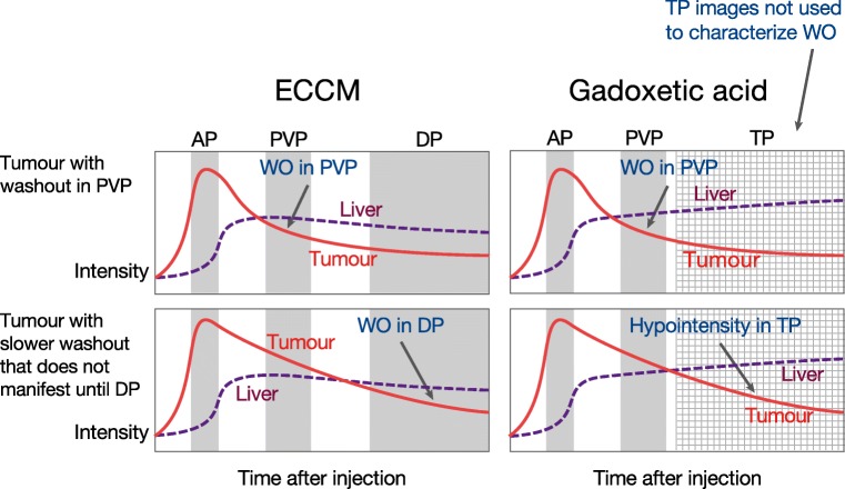
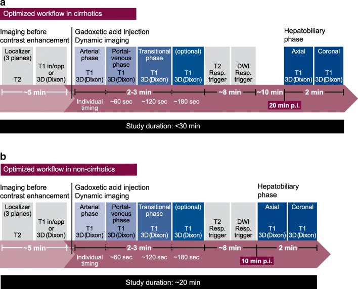
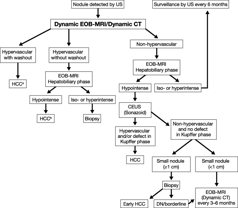
It was concluded that gadoxetic acid-enhanced MRI has higher sensitivity for the diagnosis of hepatocellular carcinoma (HCC), when compared with multidetector CT, by utilising features of hyperenhancement in the arterial phase and hypointensity in the hepatobiliary phase (HBP). Recent HCC management guidelines recognise an increasing role for gadoxetic acid-enhanced MRI in early diagnosis and monitoring post-resection. Additional research is needed to define the role of HBP in predicting microvascular invasion, to better define washout during the transitional phase in gadoxetic acid-enhanced MRI for HCC diagnosis, and to reduce the artefacts encountered in the arterial phase. Technical developments are being directed to shortening the MRI protocol for reducing time and patient discomfort and toward utilising faster imaging and non-Cartesian free-breathing approaches that have the potential to improve multiphasic dynamic imaging.
• Gadoxetic acid-enhanced MRI provides higher diagnostic sensitivity than CT for diagnosing HCC. • Gadoxetic acid-enhanced MRI has roles in early-HCC diagnosis and monitoring post-resection response. • Faster imaging and free-breathing approaches have potential to improve multiphasic dynamic imaging.
2017 年 10 月在瑞士巴塞尔举行的第八届国际肝脏磁共振成像(MRI)论坛汇聚了来自世界各地的临床和学术放射科医生,共同探讨了自 2013 年上一届论坛以来在钆塞酸增强肝脏 MRI 领域的发展和关键问题的共识。
论坛详细讨论了肝脏 MRI 的两个主要主题:在肝硬化患者中使用钆塞酸进行对比增强 MRI 和钆塞酸增强肝脏 MRI 的技术性能,这既是机遇也是挑战。本文总结了论坛上专家演讲和代表对共识声明的投票。
结论是,与多排 CT 相比,钆塞酸增强 MRI 在动脉期的高强化和肝胆期(HBP)的低信号特征上,对肝细胞癌(HCC)的诊断具有更高的敏感性。最近的 HCC 管理指南承认,钆塞酸增强 MRI 在早期诊断和监测术后方面的作用越来越大。需要进一步的研究来确定 HBP 在预测微血管侵犯中的作用,更好地定义 HCC 诊断中钆塞酸增强 MRI 过渡阶段的洗脱,以及减少动脉期遇到的伪影。技术发展旨在缩短 MRI 方案,以减少时间和患者不适,并利用更快的成像和非笛卡尔自由呼吸方法,有可能改善多期动态成像。
钆塞酸增强 MRI 提供比 CT 更高的诊断 HCC 的敏感性。
钆塞酸增强 MRI 在早期 HCC 诊断和监测术后反应中具有作用。
更快的成像和自由呼吸方法有可能改善多期动态成像。