Department of Biochemistry, University of Geneva, Geneva, Switzerland.
Cell Biology and Biophysics Unit, European Molecular Biology Laboratory (EMBL), Heidelberg, Germany.
Elife. 2019 Aug 6;8:e44215. doi: 10.7554/eLife.44215.
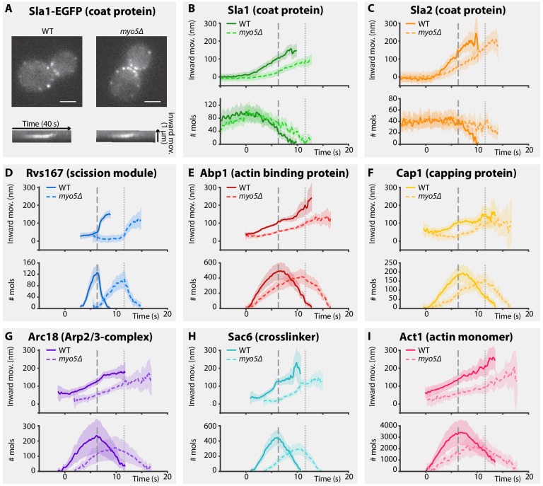
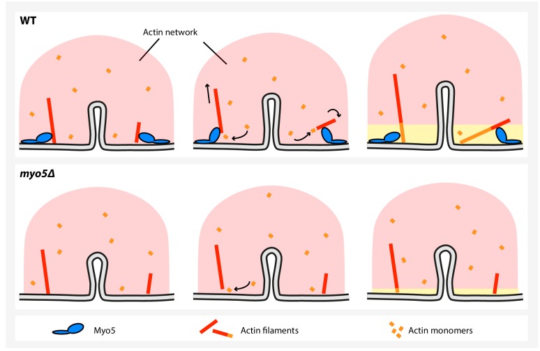
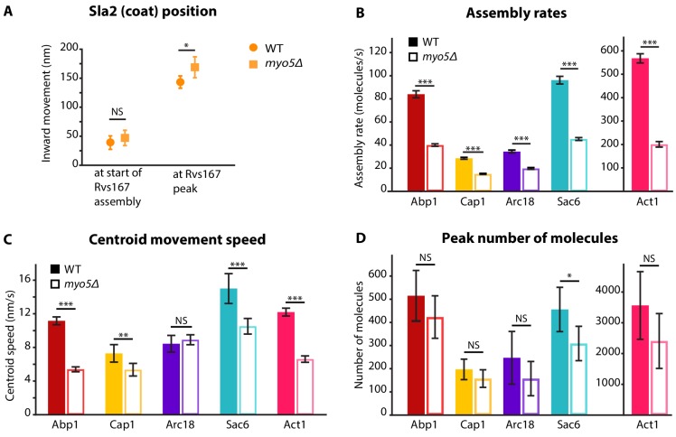
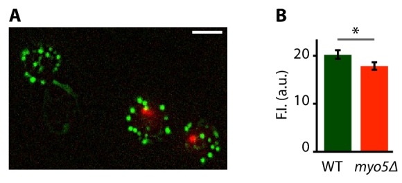
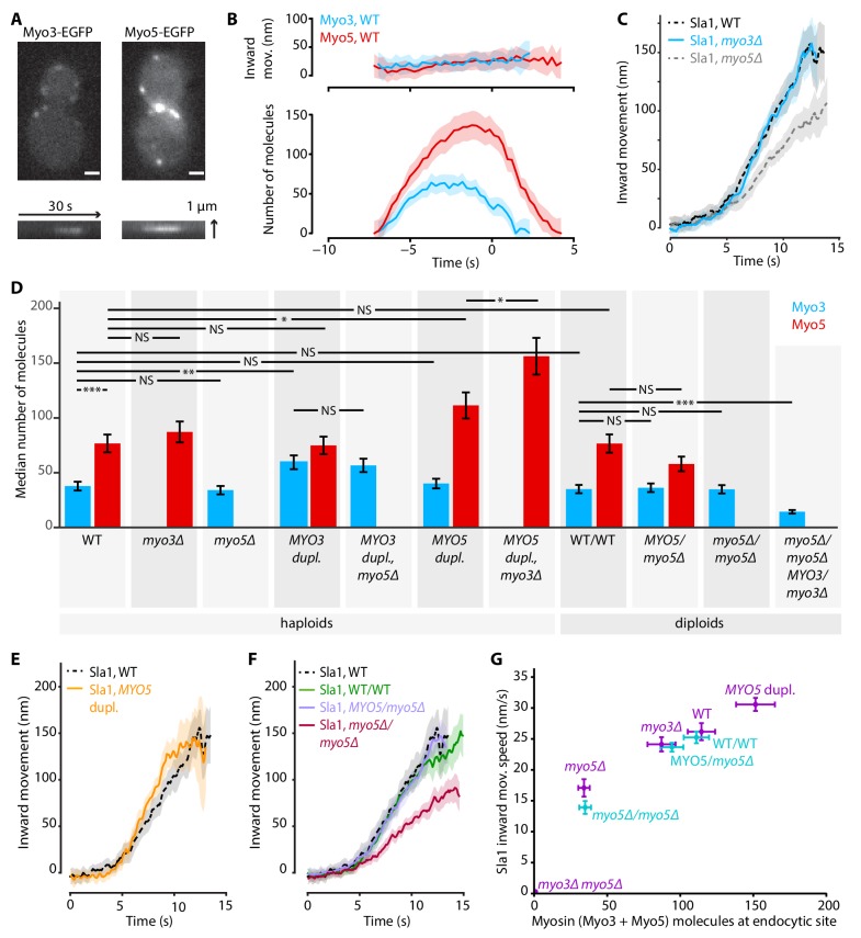
Clathrin-mediated endocytosis in budding yeast requires the formation of a dynamic actin network that produces the force to invaginate the plasma membrane against the intracellular turgor pressure. The type-I myosins Myo3 and Myo5 are important for endocytic membrane reshaping, but mechanistic details of their function remain scarce. Here, we studied the function of Myo3 and Myo5 during endocytosis using quantitative live-cell imaging and genetic perturbations. We show that the type-I myosins promote, in a dose-dependent way, the growth and expansion of the actin network, which controls the speed of membrane and coat internalization. We found that this myosin-activity is independent of the actin nucleation promoting activity of myosins, and cannot be compensated for by increasing actin nucleation. Our results suggest a new mechanism for type-I myosins to produce force by promoting actin filament polymerization.
网格蛋白介导的内吞作用在出芽酵母中需要形成一个动态的肌动蛋白网络,该网络产生的力可以抵抗细胞内的膨压向内质膜内陷。I 型肌球蛋白 Myo3 和 Myo5 对于内吞作用中膜的重塑很重要,但它们功能的机制细节仍然很少。在这里,我们使用定量活细胞成像和遗传扰动研究了 Myo3 和 Myo5 在胞吞作用中的功能。我们表明,I 型肌球蛋白以剂量依赖的方式促进肌动蛋白网络的生长和扩展,从而控制膜和衣被内化的速度。我们发现,这种肌球蛋白活性不依赖于肌球蛋白的肌动蛋白成核促进活性,并且不能通过增加肌动蛋白成核来补偿。我们的结果表明,I 型肌球蛋白通过促进肌动蛋白丝聚合产生力的一种新机制。