Department of Cardiology, Huiqiao Medical Center, Nanfang Hospital, Southern Medical University, Guangzhou, 510515, China; Department of Cardiology, Shunde Hospital, Southern Medical University (The First People's Hospital of Shunde Foshan), Foshan, 528308, Guangdong, China.
Department of Emergency Medicine, Nanfang Hospital, Southern Medical University, Guangzhou, 510515, China.
Redox Biol. 2019 Sep;26:101287. doi: 10.1016/j.redox.2019.101287. Epub 2019 Jul 27.
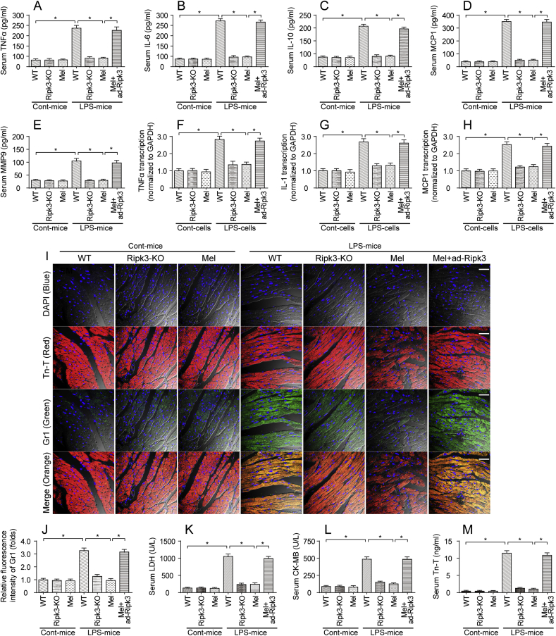
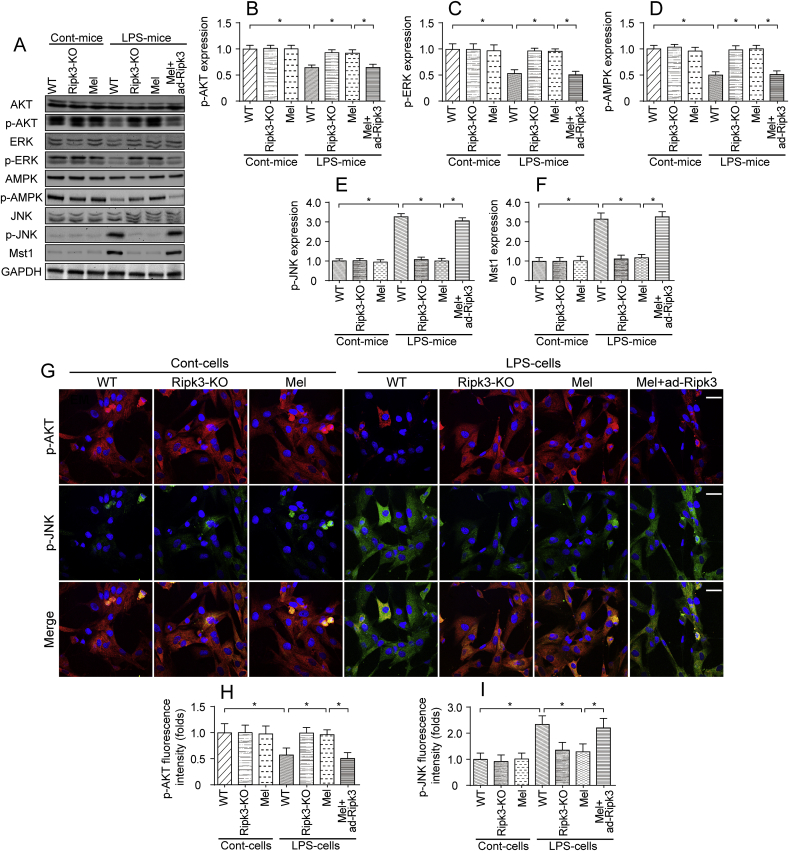
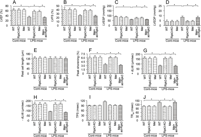
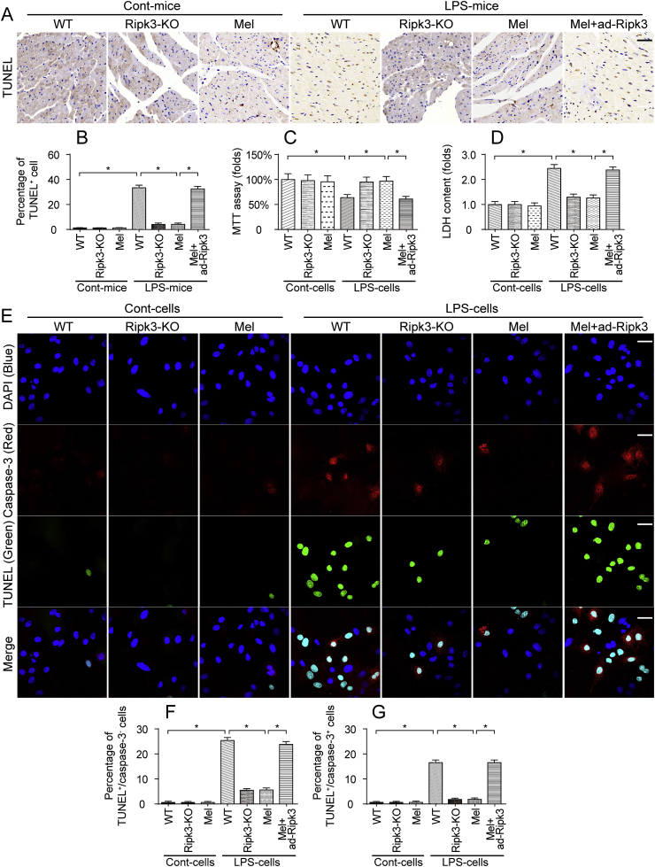
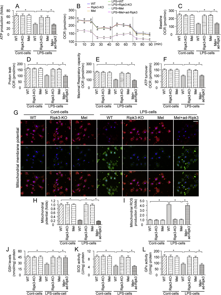
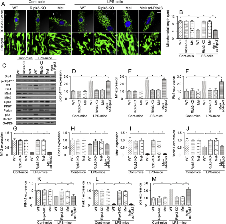
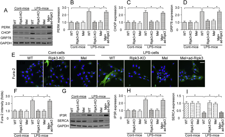
The basic pathophysiological mechanisms underlying septic cardiomyopathy have not yet been completely clarified. Disease-specific treatments are lacking, and care is still based on supportive modalities. The aim of our study was to assess the protective effects of melatonin on septic cardiomyopathy, with a focus on the interactions between receptor-interacting protein kinase 3 (Ripk3), the mitochondria, endoplasmic reticulum (ER) and cytoskeletal degradation in cardiomyocytes. Ripk3 expression was increased in heart samples challenged with LPS, followed by myocardial inflammation, cardiac dysfunction, myocardial breakdown and cardiomyocyte death. The melatonin treatment attenuated septic myocardial injury in a comparable manner to the genetic depletion of Ripk3. Molecular investigations revealed that Ripk3 intimately regulated mitochondrial function, ER stress, cytoskeletal homeostasis and cardioprotective signaling pathways. Melatonin-mediated inhibition of Ripk3 improved mitochondrial bioenergetics, reduced mitochondria-initiated oxidative damage, sustained mitochondrial dynamics, ameliorated ER stress, normalized calcium recycling, and activated cardioprotective signaling pathways (including AKT, ERK and AMPK) in cardiomyocytes. Interestingly, Ripk3 overexpression mediated resistance to melatonin therapy following the infection of LPS-treated hearts with an adenovirus expressing Ripk3. Altogether, our findings identify Ripk3 upregulation as a novel risk factor for the development of sepsis-related myocardial injury, and melatonin restores the physiological functions of the mitochondria, ER, contractile cytoskeleton and cardioprotective signaling pathways. Additionally, our data also reveal a new, potentially therapeutic mechanism by which melatonin protects the heart from sepsis-mediated dysfunction, possibly by targeting Ripk3.
脓毒症性心肌病的基本病理生理机制尚未完全阐明。目前缺乏针对该病的特异性治疗方法,治疗仍以支持性治疗为主。本研究旨在评估褪黑素对脓毒症性心肌病的保护作用,重点关注受体相互作用蛋白激酶 3(Ripk3)、线粒体、内质网(ER)和肌节细胞骨架降解之间的相互作用。LPS 刺激后的心脏样本中 Ripk3 表达增加,随后发生心肌炎症、心功能障碍、心肌破裂和心肌细胞死亡。褪黑素治疗以与 Ripk3 基因耗竭相当的方式减轻脓毒性心肌损伤。分子研究表明,Ripk3 密切调节线粒体功能、ER 应激、细胞骨架动态平衡和心脏保护信号通路。褪黑素介导的 Ripk3 抑制可改善线粒体生物能学,减少线粒体引发的氧化损伤,维持线粒体动力学,减轻 ER 应激,使钙循环正常化,并激活心肌细胞中的心脏保护信号通路(包括 AKT、ERK 和 AMPK)。有趣的是,Ripk3 过表达介导了 LPS 处理的心脏感染表达 Ripk3 的腺病毒后对褪黑素治疗的耐药性。总之,我们的研究结果表明 Ripk3 上调是脓毒症相关心肌损伤发展的一个新的危险因素,而褪黑素恢复了线粒体、ER、收缩细胞骨架和心脏保护信号通路的生理功能。此外,我们的数据还揭示了褪黑素通过靶向 Ripk3 保护心脏免受脓毒症介导的功能障碍的新的、潜在的治疗机制。