Department of Geriatrics, Xijing Hospital, The Fourth Military Medical University, Xi'an, China.
Department of Thoracic Surgery, Sixth Medical Center of PLA General Hospital, Beijing, China.
Front Endocrinol (Lausanne). 2020 Apr 16;11:162. doi: 10.3389/fendo.2020.00162. eCollection 2020.
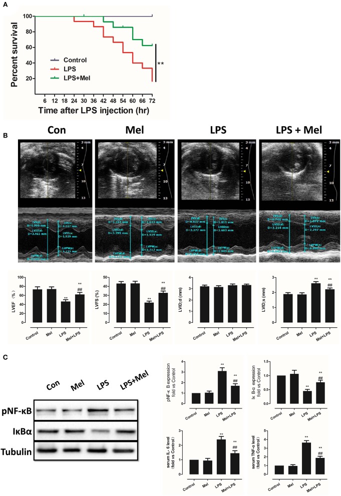
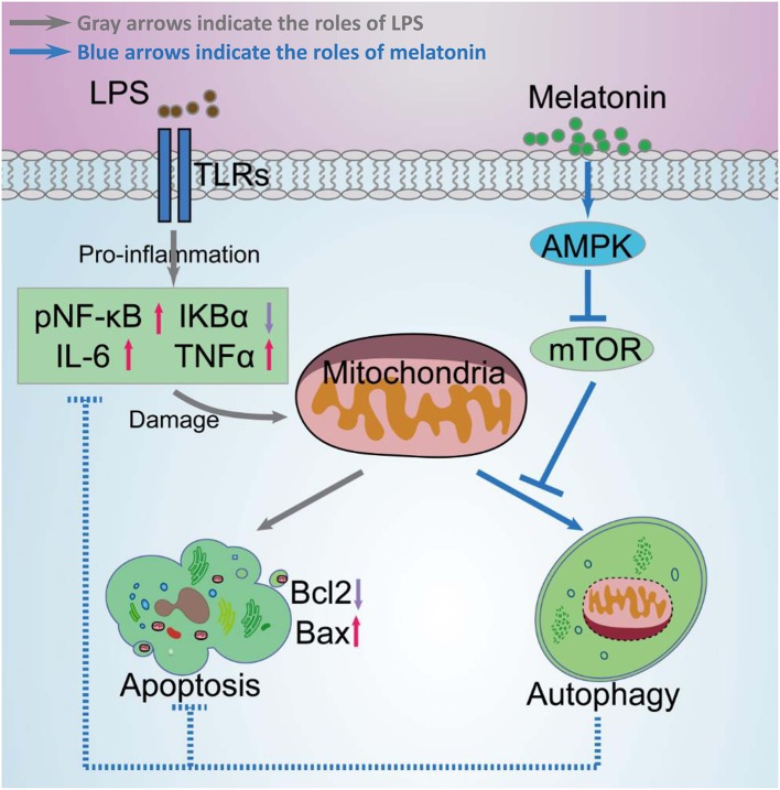
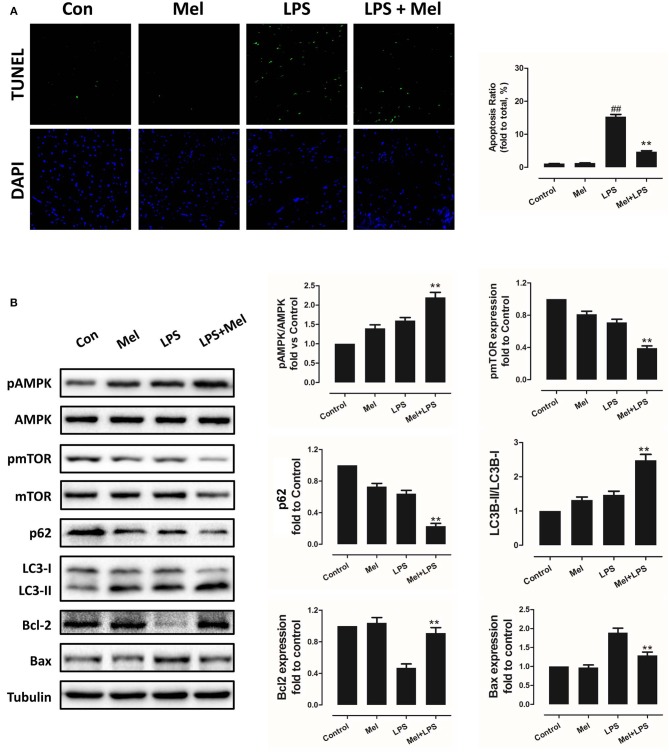
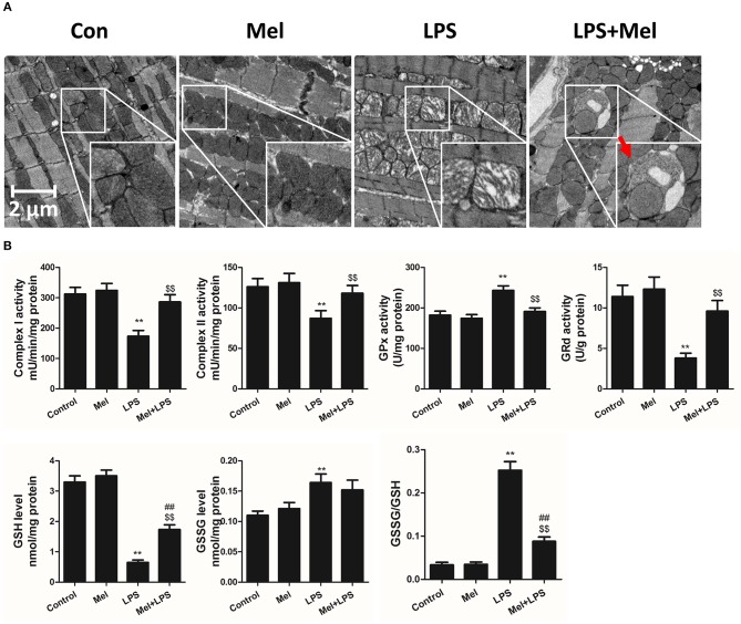
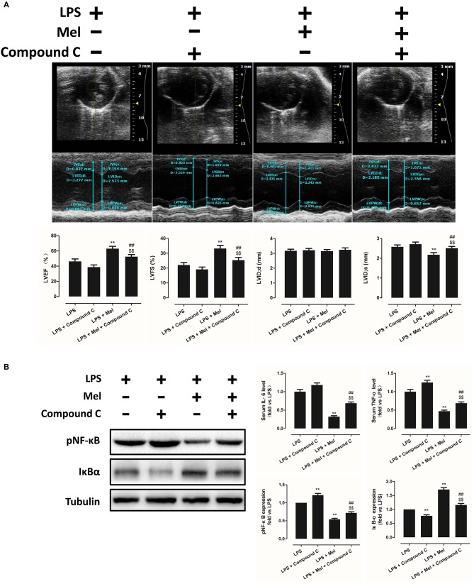
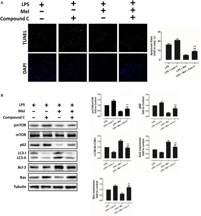
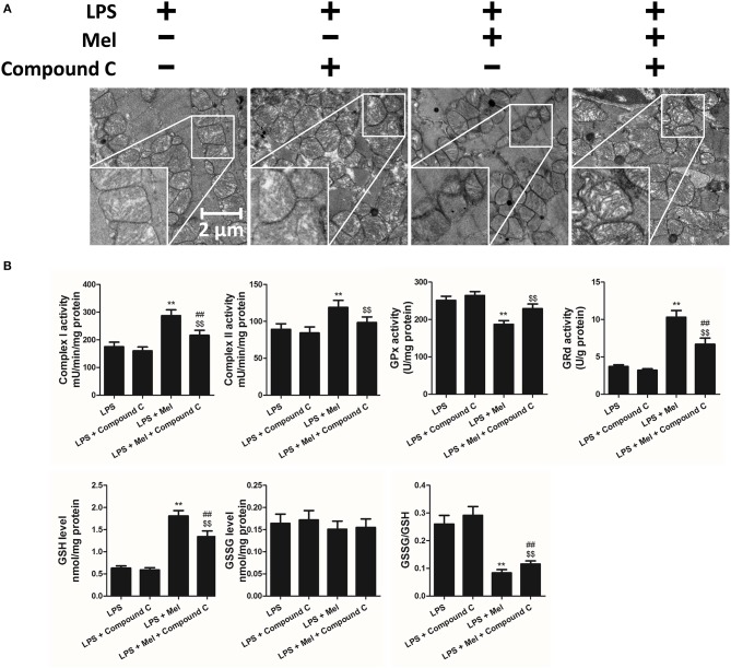
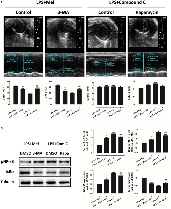
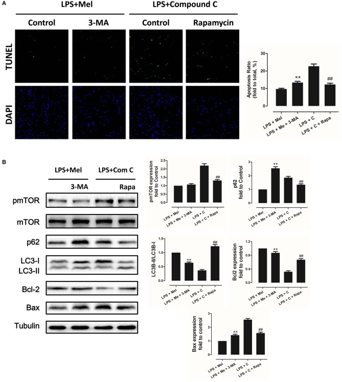
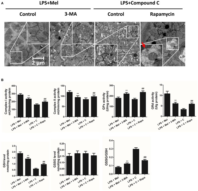
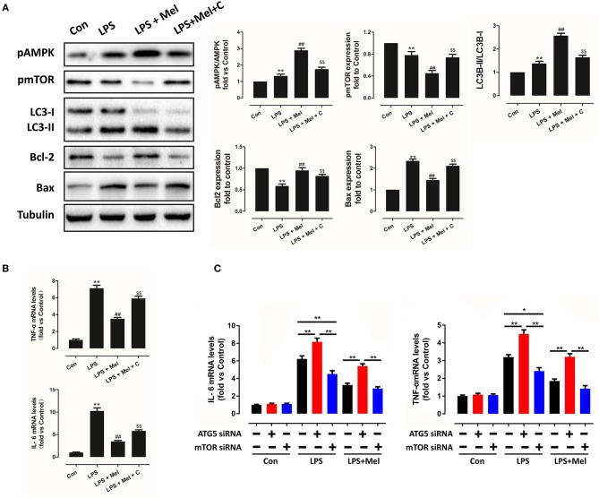
Melatonin is an indolamine secreted by the pineal gland, as well as most of the organs and tissues. In addition to regulating circadian biology, studies have confirmed the multiple pharmacological effects of melatonin. Melatonin provides a strong defense against septic myocardial injury. However, the underlying mechanism has not been fully described. In this study, we investigated the protective effects of melatonin against lipopolysaccharide (LPS)-induced myocardial injury as well as the mechanisms involved. Mice were intraperitoneally injected with LPS to induce a septic myocardial injury model or an LPS shock model, depending on the dose of LPS. Melatonin was given (20 mg/kg/day, via intraperitoneal injection) for a week prior to LPS insult. 6 h after LPS injection, echocardiographic analysis, TUNEL staining, transmission electron microscopy (TEM), western blot, quantitative real-time PCR and ELISA were used to investigate the protective effects of melatonin against LPS induced myocardial injury. AMPK inhibitor, autophagy activator and inhibitor, siRNAs were used for further validation. Survival test showed that melatonin significantly increased the survival rate after LPS-induced shock. In the sepsis model, melatonin markedly ameliorated myocardial dysfunction, decreased the release of inflammatory cytokines, activated AMP-activated protein kinase (AMPK), improved mitochondrial function, and activated autophagy. To confirm whether the protection of melatonin was mediated by AMPK and autophagy, Compound C, an AMPK inhibitor; 3-MA, an autophagy inhibitor; and Rapamycin (Rapa), an autophagy activator, were used in this study. AMPK inhibition down-regulated autophagy, abolished protection of melatonin, as indicated by significantly decreased cardiac function, increased inflammation and damaged mitochondrial function. Furthermore, autophagy inhibition by 3-MA significantly impaired the protective effects of melatonin, whereas autophagy activation by Rapa reversed LPS + Compound C induced myocardial injury. In addition, studies further confirmed the protection of melatonin against LPS-induced myocardial injury and the mechanisms involving AMPK-mediated autophagy signaling. In summary, our results demonstrated that melatonin protects against LPS-induced septic myocardial injury by activating AMPK mediated autophagy pathway.
褪黑素是由松果体分泌的一种吲哚胺,也是大多数器官和组织分泌的物质。除了调节昼夜生物节律,研究已经证实褪黑素具有多种药理学作用。褪黑素为脓毒症心肌损伤提供了强大的防御。然而,其潜在的机制尚未完全描述。在这项研究中,我们研究了褪黑素对脂多糖(LPS)诱导的心肌损伤的保护作用及其机制。 我们通过腹腔内注射 LPS 诱导脓毒症心肌损伤模型或 LPS 休克模型,具体取决于 LPS 的剂量。在 LPS 损伤前一周,通过腹腔内注射给予褪黑素(20mg/kg/天)。 LPS 注射后 6 小时,通过超声心动图分析、TUNEL 染色、透射电子显微镜(TEM)、western blot、实时定量 PCR 和 ELISA 检测褪黑素对 LPS 诱导的心肌损伤的保护作用。使用 AMPK 抑制剂、自噬激活剂和抑制剂、siRNA 进行进一步验证。 生存试验表明,褪黑素显著提高 LPS 诱导休克后的存活率。在脓毒症模型中,褪黑素显著改善心肌功能障碍,减少炎症细胞因子的释放,激活 AMP 激活的蛋白激酶(AMPK),改善线粒体功能,并激活自噬。为了证实褪黑素的保护作用是否由 AMPK 和自噬介导,本研究使用了 AMPK 抑制剂 Compound C、自噬抑制剂 3-MA 和自噬激活剂 Rapamycin(Rapa)。AMPK 抑制下调自噬,显著降低了褪黑素的保护作用,表现为心功能明显下降、炎症增加和线粒体功能受损。此外,3-MA 抑制自噬显著削弱了褪黑素的保护作用,而 Rapa 激活自噬则逆转了 LPS+Compound C 诱导的心肌损伤。此外,进一步证实了褪黑素对 LPS 诱导的心肌损伤的保护作用及其涉及 AMPK 介导的自噬信号通路的机制。 综上所述,我们的研究结果表明,褪黑素通过激活 AMPK 介导的自噬通路来保护 LPS 诱导的脓毒症心肌损伤。