Zhao Zhaoning, Zhu Pingjun, Lou Yue, Hou Ruoyu, Sun Heqiang, Du Yingzhen, Xu Guogang
Health Management Institute, The Second Medical Center & National Clinical Research Center for Geriatric Diseases, Chinese PLA General Hospital, Beijing 100853, China.
Chinese PLA General Hospital, Medical School of Chinese PLA, Beijing 100853, China.
Int J Med Sci. 2025 Jan 1;22(1):71-86. doi: 10.7150/ijms.104932. eCollection 2025.
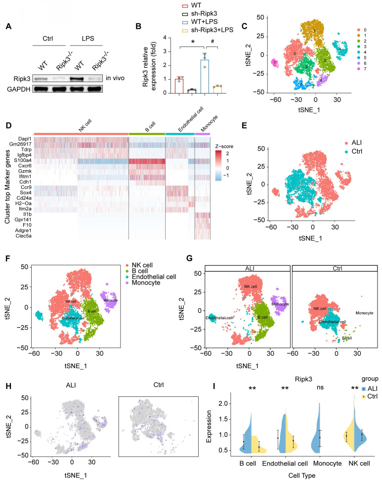
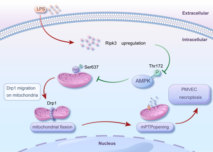
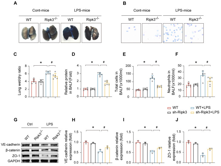
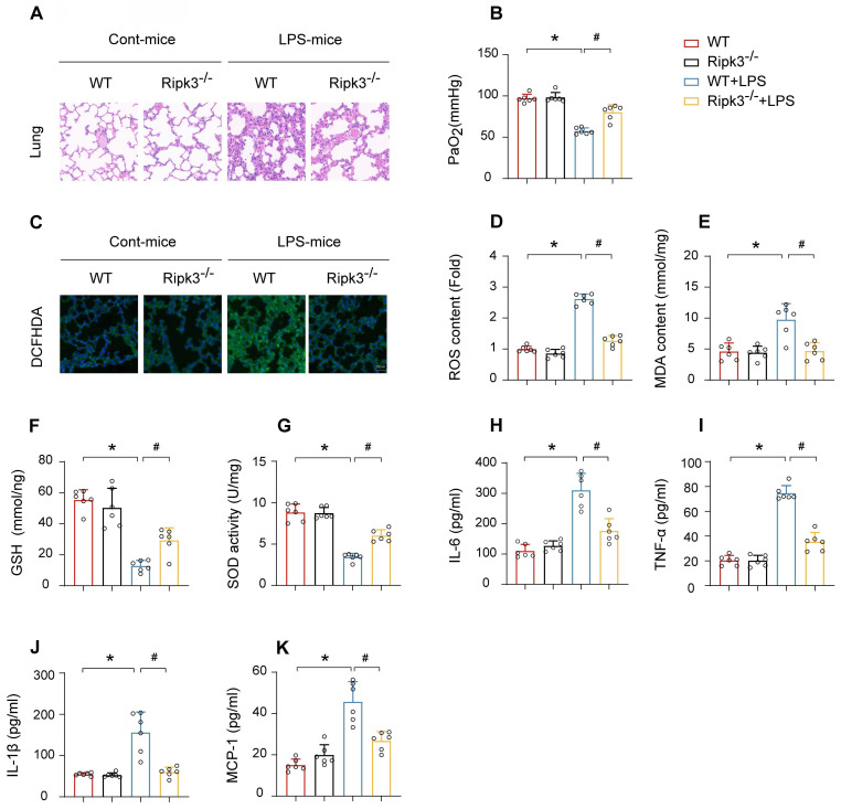
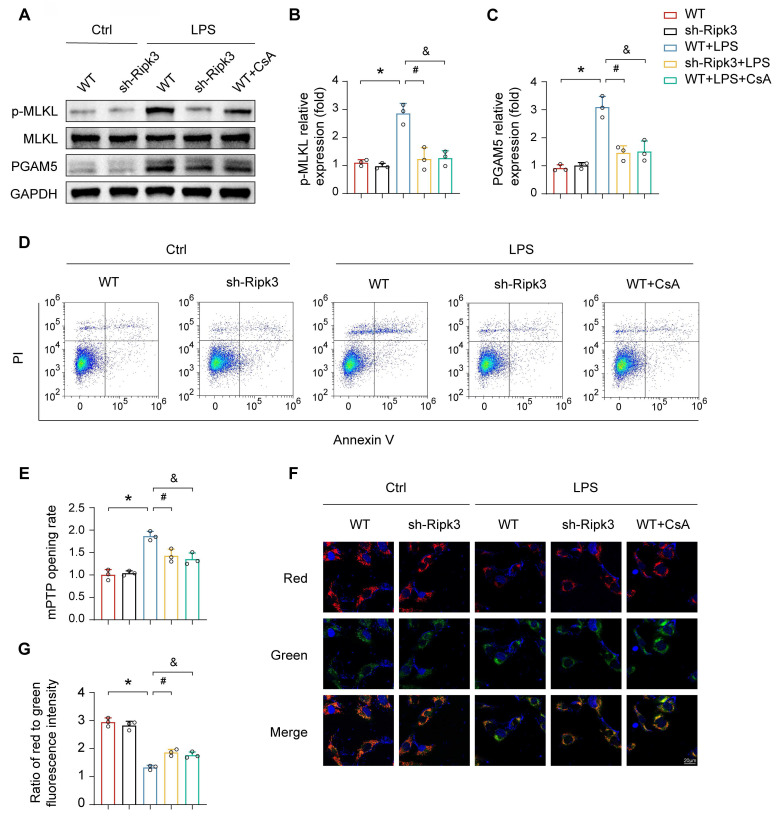
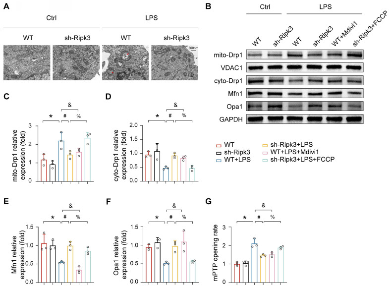
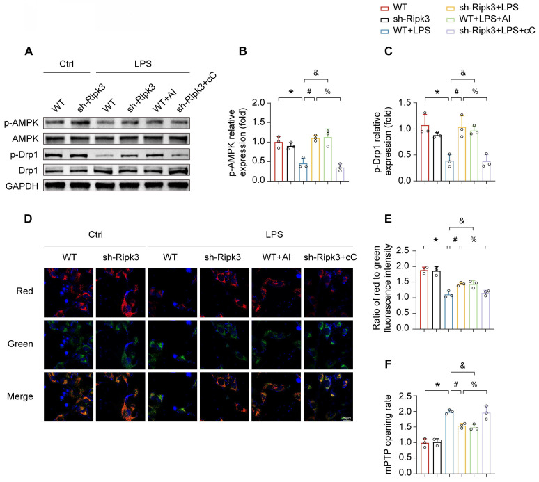
Receptor-interacting protein 3 (Ripk3) plays a crucial part in acute lung injury (ALI) by regulating inflammation-induced endothelial damage in the lung tissue. The precise mechanisms through which Ripk3 contributes to the endothelial injury in ALI still remain uncertain. In the current research, we employed Ripk3-deficient (Ripk3) mice to examine the role of Ripk3 in ALI progression, focusing on its effects on endothelial cells (ECs), mitochondrial damage and necroptosis. Our study observed significant Ripk3 upregulation in lipopolysaccharide- (LPS-) treated lung tissues, as well as in murine pulmonary microvascular endothelial cells (PMVECs). Ripk3 deletion improved lung tissue morphology, reduced inflammation, oxidative stress and endothelial dysfunction under LPS challenge. It also mitigated LPS-induced necroptosis and mitochondrial damage in PMVECs. Ripk3 upregulation suppressed the AMP-activated protein kinase (AMPK) pathway and activated Drp1-mediated mitochondrial fission, increasing mitochondrial permeability transition pore (mPTP) opening and PMVEC necroptosis. Conversely, Ripk3 deletion activated the AMPK/Drp1-mitochondrial fission pathway, preventing mPTP opening and PMVEC necroptosis in ALI. These findings demonstrated that Ripk3 promotes necroptosis through the AMPK/Drp1/mPTP opening pathway, identifying a potential therapeutic target for ALI treatment.
受体相互作用蛋白3(Ripk3)通过调节肺组织中炎症诱导的内皮损伤,在急性肺损伤(ALI)中起关键作用。Ripk3导致ALI中内皮损伤的确切机制仍不清楚。在当前的研究中,我们使用Ripk3基因敲除(Ripk3 -/-)小鼠来研究Ripk3在ALI进展中的作用,重点关注其对内皮细胞(ECs)、线粒体损伤和坏死性凋亡的影响。我们的研究观察到,在脂多糖(LPS)处理的肺组织以及小鼠肺微血管内皮细胞(PMVECs)中,Ripk3显著上调。在LPS刺激下,Ripk3基因敲除改善了肺组织形态,减轻了炎症、氧化应激和内皮功能障碍。它还减轻了LPS诱导的PMVECs坏死性凋亡和线粒体损伤。Ripk3上调抑制了AMP激活的蛋白激酶(AMPK)途径,并激活了Drp1介导的线粒体分裂,增加了线粒体通透性转换孔(mPTP)的开放和PMVECs坏死性凋亡。相反,Ripk3基因敲除激活了AMPK/Drp1 - 线粒体分裂途径,在ALI中防止了mPTP开放和PMVECs坏死性凋亡。这些发现表明,Ripk3通过AMPK/Drp1/mPTP开放途径促进坏死性凋亡,确定了一个用于ALI治疗的潜在靶点。