Tan Ying, Wu Xunhua, Chen Jing, Kong Lingyu, Qian Zhaoxin
Health Management Center, Xiangya Hospital, Central South University, Changsha, China.
Department of Radiology, Central Xiangya Hospital, South University, Changsha, China.
Front Psychol. 2019 Jul 25;10:1700. doi: 10.3389/fpsyg.2019.01700. eCollection 2019.
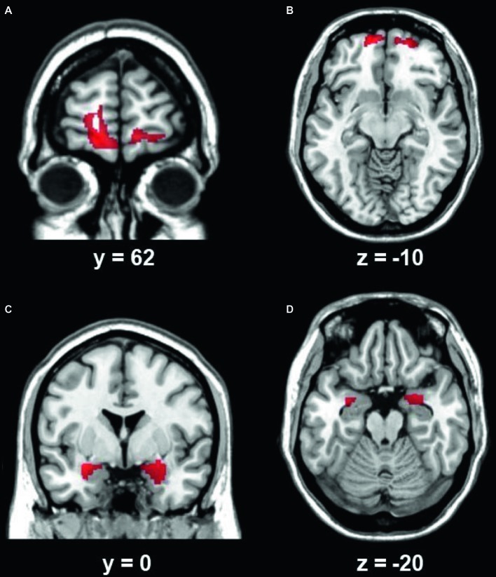
Featuring a burning sensation in the tongue or other oral sites in the absence of observable lesions or laboratory findings, burning mouth syndrome (BMS) is a chronic intraoral pain disorder, which is one of the most common medically unexplained oral symptoms/syndromes. Previous studies have suggested that brain changes are involved in BMS; however, the small number of participants in these studies limited the conclusions that could be drawn. The present study aimed to further elucidate the brain anatomical and functional changes in BMS with a relatively large sample. Fifty-three patients (26 BMS patients and 27 gender- and age-matched controls) were recruited. Demographic information was collected interviews. Visual analogue scale (VAS), anxiety, and depression scale were administered. Participants underwent an MRI scan (including one high-resolution structural scan, one diffusion tensor image, and one session of resting state scan) on the same day. The results showed that BMS patients had higher depression and anxiety levels than controls. BMS patients showed lower gray matter volume (GMV) in the bilateral ventromedial prefrontal cortex (VMPFC) and increased functional connectivity between this region and the bilateral amygdala. Region of interest (ROI) analysis suggested that the functional connectivity between the bilateral VMPFC and amygdala correlated with the years of BMS illness in patients. The brain measures could predict the years of symptoms in the BMS group. These results suggest A potential neuromarker for the diagnosis and treatment of BMS.
灼口综合征(BMS)的特征是在舌头或其他口腔部位有烧灼感,而无明显病变或实验室检查结果,它是一种慢性口腔内疼痛疾病,是最常见的医学上无法解释的口腔症状/综合征之一。先前的研究表明,大脑变化与灼口综合征有关;然而,这些研究中的参与者数量较少,限制了所能得出的结论。本研究旨在通过相对较大的样本进一步阐明灼口综合征患者大脑的解剖和功能变化。招募了53名患者(26名灼口综合征患者和27名性别及年龄匹配的对照者)。通过访谈收集人口统计学信息。使用视觉模拟量表(VAS)、焦虑和抑郁量表进行评估。参与者在同一天接受了MRI扫描(包括一次高分辨率结构扫描、一次弥散张量成像和一次静息态扫描)。结果显示,灼口综合征患者的抑郁和焦虑水平高于对照组。灼口综合征患者双侧腹内侧前额叶皮质(VMPFC)的灰质体积(GMV)较低,且该区域与双侧杏仁核之间的功能连接增加。感兴趣区(ROI)分析表明,双侧VMPFC与杏仁核之间的功能连接与患者灼口综合征的患病年限相关。大脑测量指标可以预测灼口综合征组的症状持续年限。这些结果提示了一种用于灼口综合征诊断和治疗的潜在神经标志物。