G.E.R.N. Tissue Replacement, Regeneration & Neogenesis, Department of Orthopedics and Trauma Surgery, Medical Center - Albert-Ludwigs-University of Freiburg, Faculty of Medicine, Albert-Ludwigs-University of Freiburg, Freiburg im Breisgau, Germany.
Schoen Clinic Munich Harlaching, Teaching Hospital of Paracelsus Medical University Salzburg, Salzburg, Austria.
J Orthop Surg Res. 2019 Aug 13;14(1):256. doi: 10.1186/s13018-019-1308-5.
There are many studies on osteoarthritis, but only a few studies deal with human arthrosis, comparing the mechanical properties of healthy and diseased samples. In most of these studies, only isolated areas of the tibia are examined. There is currently only one study investigating the complete mapping of cartilage tissue but not the difference between instantaneous modulus (IM) in healthy and diseased samples. The aim of this study is to investigate the relationship between the biomechanical and histological changes of articular cartilage in the pathogenesis of osteoarthritis.
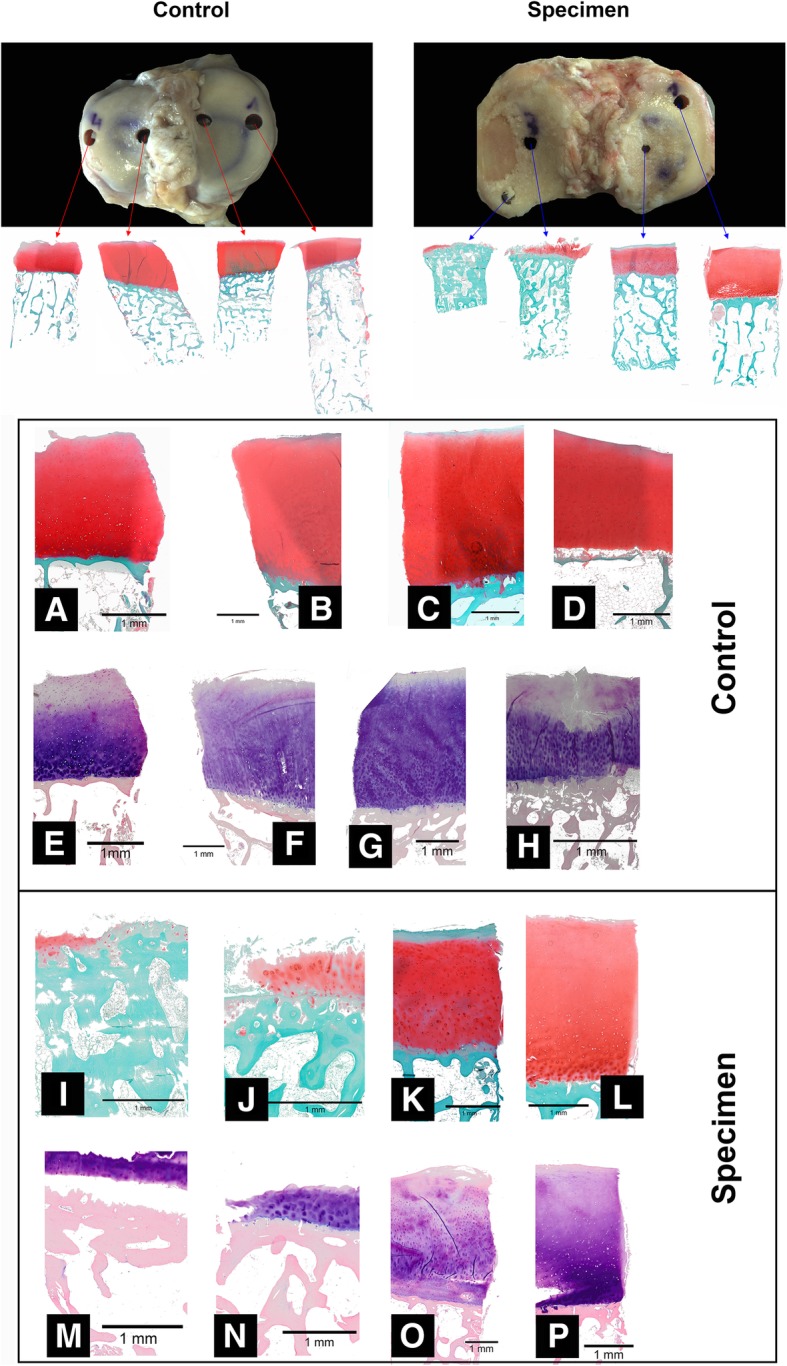
The study compared 25 tibiae with medial gonarthrosis and 13 healthy controls. The IM was determined by automated indentation mapping using a Mach-1 V500css testing machine. A grid was projected over the sample and stored so that all measurements could be taken at the same positions (100 ± 29 positions across the tibiae). This grid was then used to perform the thickness measurement using the needle method. Samples were then taken for histological examinations using a hollow milling machine. Then Giemsa and Safranin O staining were performed. In order to determine the degree of arthrosis according to histological criteria, the assessment was made with regard to Osteoarthritis Research Society International (OARSI) and AHO scores.
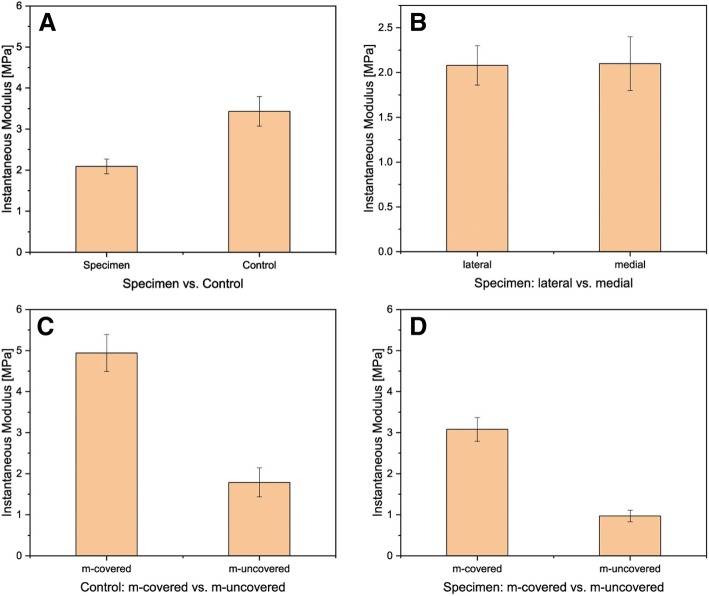
A significant difference (p < 0.05) could be observed in the measured IM between the controls with 3.43 ± 0.36 MPa and the samples with 2.09 ± 0.18 MPa. In addition, there was a significant difference in IM in terms of meniscus-covered and meniscus-uncovered areas. The difference in cartilage thickness between 2.25 ± 0.11 mm controls and 2.0 ± 0.07 mm samples was highly significant with p < 0.001. With regard to the OARSI and AHO scores, the samples differed significantly from the controls. The OARSI and AHO scores showed a significant difference between meniscus-covered and meniscus-uncovered areas.
The controls showed significantly better viscoelastic behavior than the arthrotic samples in the measured IM. The measured biomechanical values showed a direct correlation between histological changes and altered biomechanics in gonarthrosis.
有许多关于骨关节炎的研究,但只有少数研究涉及人类关节病,比较健康和患病样本的机械性能。在这些研究中,大多数只检查胫骨的孤立区域。目前只有一项研究调查软骨组织的完整图谱,但没有研究健康和患病样本之间的瞬时模量(IM)差异。本研究旨在探讨关节软骨在骨关节炎发病机制中的生物力学和组织学变化之间的关系。
该研究比较了 25 例内侧膝关节骨关节炎和 13 例健康对照的胫骨。使用 Mach-1 V500css 试验机通过自动压痕映射确定 IM。将网格投影到样本上并存储,以便可以在相同位置(胫骨上 100 ± 29 个位置)进行所有测量。然后使用该网格通过针测法进行厚度测量。使用空心铣床采集样本进行组织学检查。然后进行 Giemsa 和 Safranin O 染色。为了根据组织学标准确定关节炎的程度,根据骨关节炎研究协会国际(OARSI)和 AHO 评分进行评估。
在对照组的 3.43 ± 0.36 MPa 和样本的 2.09 ± 0.18 MPa 之间,可观察到测量的 IM 存在显著差异(p < 0.05)。此外,在半月板覆盖和半月板未覆盖区域的 IM 也存在显著差异。2.25 ± 0.11 mm 对照组和 2.0 ± 0.07 mm 样本之间的软骨厚度差异具有高度显著性,p < 0.001。关于 OARSI 和 AHO 评分,样本与对照组有显著差异。OARSI 和 AHO 评分在半月板覆盖和半月板未覆盖区域之间存在显著差异。
对照组在测量的 IM 中表现出明显优于关节炎样本的粘弹性行为。测量的生物力学值显示出膝关节骨关节炎中组织学变化与生物力学改变之间的直接相关性。