• 文献检索
  • 文档翻译
  • 深度研究
  • 学术资讯
  • Suppr Zotero 插件Zotero 插件
  • 邀请有礼
  • 套餐&价格
  • 历史记录
应用&插件
Suppr Zotero 插件Zotero 插件浏览器插件Mac 客户端Windows 客户端微信小程序
定价
高级版会员购买积分包购买API积分包
服务
文献检索文档翻译深度研究API 文档MCP 服务
关于我们
关于 Suppr公司介绍联系我们用户协议隐私条款
关注我们

Suppr 超能文献

核心技术专利:CN118964589B侵权必究
粤ICP备2023148730 号-1Suppr @ 2026

文献检索

告别复杂PubMed语法,用中文像聊天一样搜索,搜遍4000万医学文献。AI智能推荐,让科研检索更轻松。

立即免费搜索

文件翻译

保留排版,准确专业,支持PDF/Word/PPT等文件格式,支持 12+语言互译。

免费翻译文档

深度研究

AI帮你快速写综述,25分钟生成高质量综述,智能提取关键信息,辅助科研写作。

立即免费体验

二氧化硅纳米颗粒与过氧化氢酶和人骨髓间充质干细胞的相互作用:生物物理、理论和细胞研究。

The interaction of silica nanoparticles with catalase and human mesenchymal stem cells: biophysical, theoretical and cellular studies.

机构信息

Department of Biochemistry and Biophysics, Faculty of Advanced Science and Technology, Tehran Medical Sciences, Islamic Azad University, Tehran, Iran.

Department of Medical Laboratory Technology, Health Technical College, Erbil Polytechnic University, Erbil, Kurdistan Region, Iraq.

出版信息

Int J Nanomedicine. 2019 Jul 16;14:5355-5368. doi: 10.2147/IJN.S210136. eCollection 2019.

DOI:10.2147/IJN.S210136
PMID:31409992
原文链接:https://pmc.ncbi.nlm.nih.gov/articles/PMC6643057/
Abstract

AIM

Nanoparticles (NPs) have been receiving potential interests in protein delivery and cell therapy. As a matter of fact, NPs may be used as great candidates in promoting cell therapy by catalase (CAT) delivery into high oxidative stress tissues. However, for using NPs like SiO as carriers, the interaction of NPs with proteins and mesenchymal stem cells (MSCs) should be explored in advance.

METHODS

In the present study, the interaction of SiO NPs with CAT and human MSCs (hMSCs) was explored by various spectroscopic methods (fluorescence, circular dichroism (CD), UV-visible), molecular docking and dynamics studies, and cellular (MTT, cellular morphology, cellular uptake, lactate dehydrogenase, ROS, caspase-3, flow cytometry) assays.

RESULTS

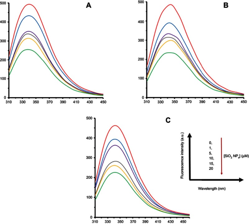
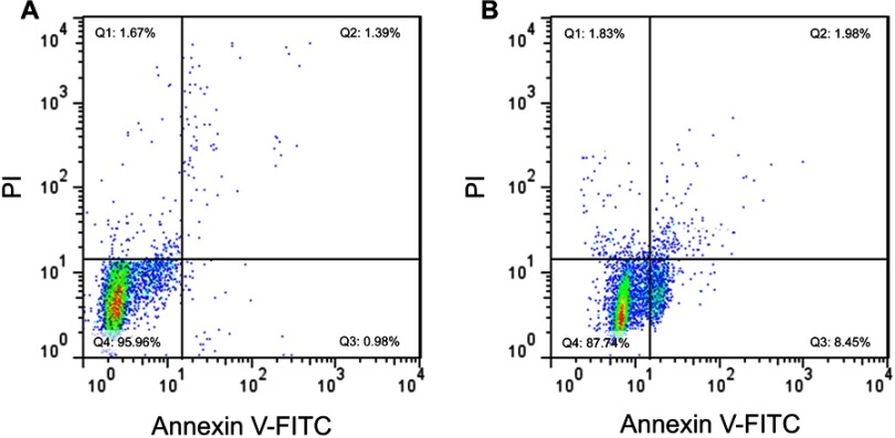
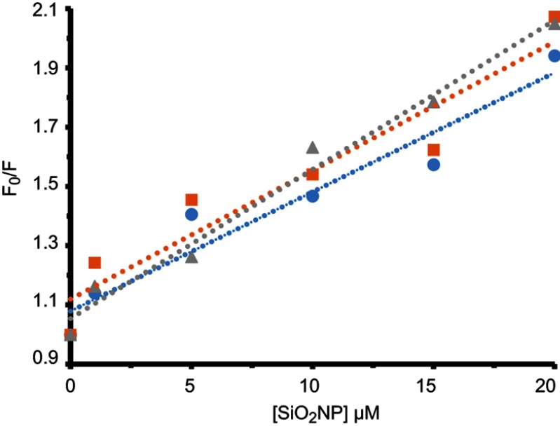
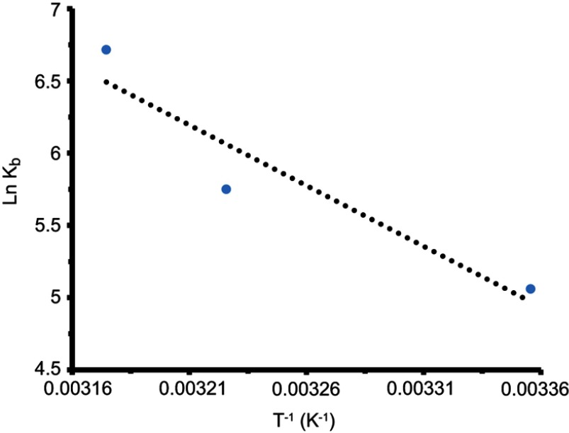
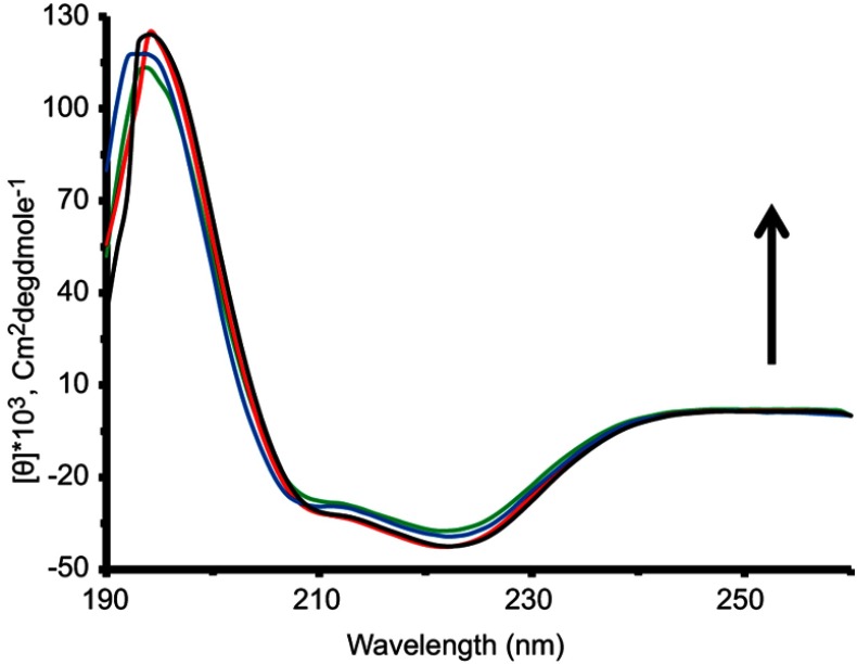
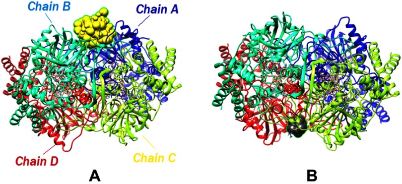
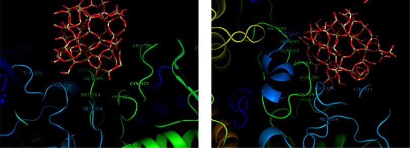
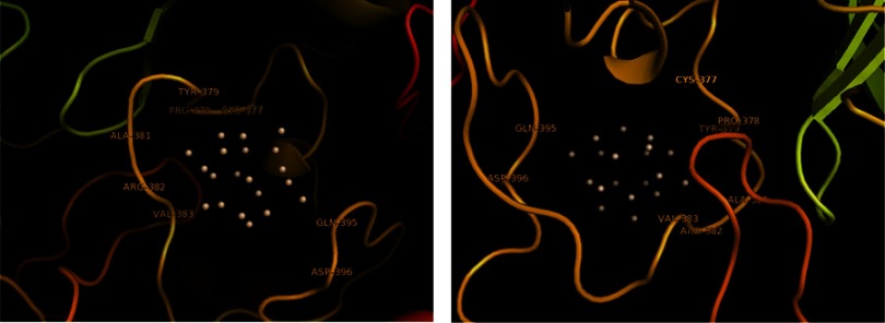
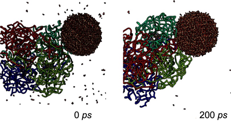
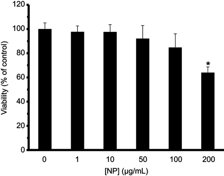
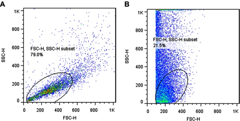
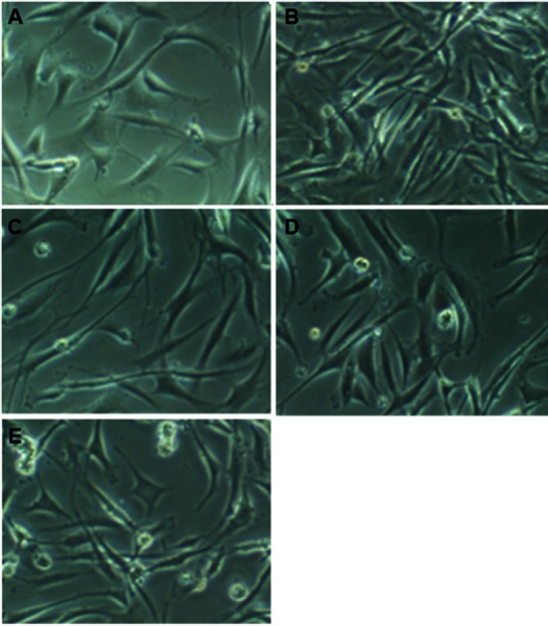
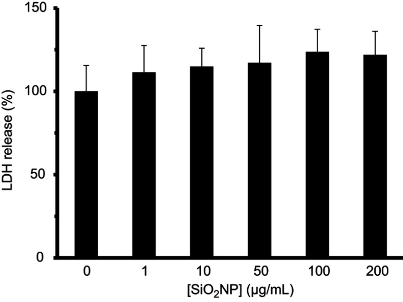
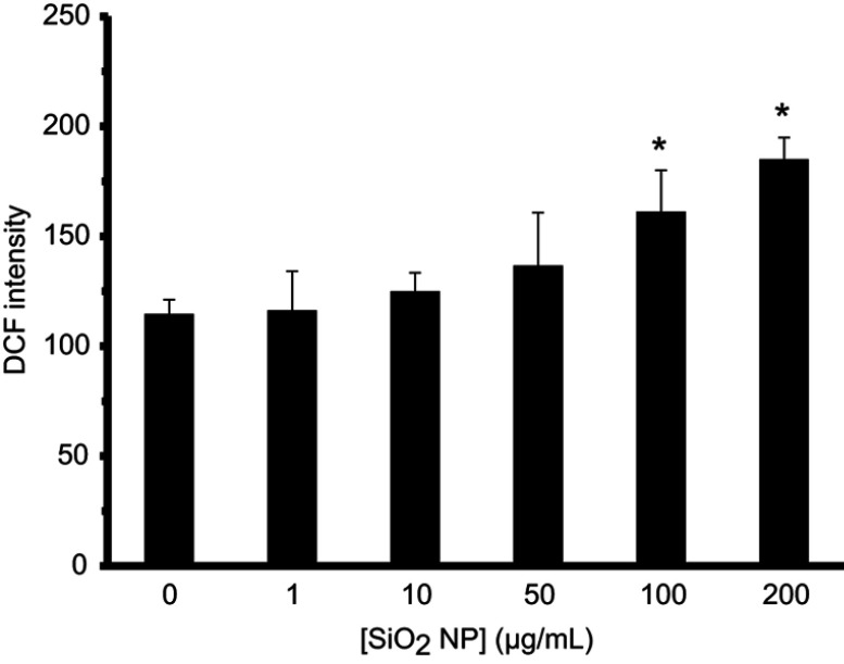
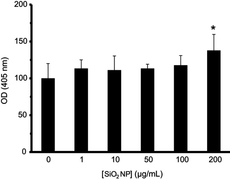
Fluorescence study displayed that both dynamic and static quenching mechanisms and hydrophobic interactions are involved in the spontaneous interaction of SiO NPs with CAT. CD spectra indicated that native structure of CAT remains stable after interaction with SiO NPs. UV-visible study also revealed that the kinetic parameters of CAT such as , and enzyme efficiency were not changed after the addition of SiO NPs. Molecular docking and dynamics studies showed that Si and SiO clusters interact with hydrophobic residues of CAT and SiO cluster causes minor changes in the CAT structure at a total simulation time of 200 ps. Cellular assays depicted that SiO NPs induce significant cell mortality, change in cellular morphology, cellular internalization, ROS elevation, and apoptosis in hMSCs at higher concentration than 100 µg/mL (170 µM).

CONCLUSION

The current results suggest that low concentrations of SiO NPs induce no substantial change or mortality against CAT and hMSCs, and potentially useful carriers in CAT delivery to hMSC.

摘要

目的

纳米粒子(NPs)在蛋白质递药和细胞治疗方面受到了广泛关注。事实上,NPs 可以作为细胞治疗的优秀候选者,通过将过氧化氢酶(CAT)递送至高氧化应激组织中来促进细胞治疗。然而,对于使用 SiO2 NPs 等作为载体,需要预先探索 NPs 与蛋白质和间充质干细胞(MSCs)的相互作用。

方法

在本研究中,通过各种光谱方法(荧光、圆二色性(CD)、紫外-可见)、分子对接和动力学研究以及细胞(MTT、细胞形态、细胞摄取、乳酸脱氢酶、ROS、caspase-3、流式细胞术)实验,研究了 SiO2 NPs 与 CAT 和人 MSCs(hMSCs)的相互作用。

结果

荧光研究表明,SiO2 NPs 与 CAT 的自发相互作用涉及动态和静态猝灭机制以及疏水相互作用。CD 光谱表明,CAT 的天然结构在与 SiO2 NPs 相互作用后保持稳定。紫外-可见研究还表明,CAT 的动力学参数,如、和酶效率,在加入 SiO2 NPs 后没有改变。分子对接和动力学研究表明,Si 和 SiO 簇与 CAT 的疏水残基相互作用,SiO 簇在总共 200 ps 的模拟时间内导致 CAT 结构的微小变化。细胞实验表明,SiO2 NPs 在高于 100 µg/mL(170 µM)的浓度下,会导致 hMSCs 发生显著的细胞死亡、细胞形态改变、细胞内吞、ROS 升高和凋亡。

结论

目前的结果表明,低浓度的 SiO2 NPs 对 CAT 和 hMSCs 没有引起实质性的变化或死亡率,并且可能是 CAT 递送至 hMSC 的有用载体。

https://cdn.ncbi.nlm.nih.gov/pmc/blobs/c53d/6643057/b730356729d6/IJN-14-5355-g0016.jpg
https://cdn.ncbi.nlm.nih.gov/pmc/blobs/c53d/6643057/53bf015ebf6a/IJN-14-5355-g0001.jpg
https://cdn.ncbi.nlm.nih.gov/pmc/blobs/c53d/6643057/7ad58860353d/IJN-14-5355-g0002.jpg
https://cdn.ncbi.nlm.nih.gov/pmc/blobs/c53d/6643057/6c7c7e5dc817/IJN-14-5355-g0003.jpg
https://cdn.ncbi.nlm.nih.gov/pmc/blobs/c53d/6643057/cc216d18f39c/IJN-14-5355-g0004.jpg
https://cdn.ncbi.nlm.nih.gov/pmc/blobs/c53d/6643057/4f2915143379/IJN-14-5355-g0005.jpg
https://cdn.ncbi.nlm.nih.gov/pmc/blobs/c53d/6643057/d88a9c726305/IJN-14-5355-g0006.jpg
https://cdn.ncbi.nlm.nih.gov/pmc/blobs/c53d/6643057/5350c80e8088/IJN-14-5355-g0007.jpg
https://cdn.ncbi.nlm.nih.gov/pmc/blobs/c53d/6643057/36dbb45af3fb/IJN-14-5355-g0008.jpg
https://cdn.ncbi.nlm.nih.gov/pmc/blobs/c53d/6643057/c6d0eab5763b/IJN-14-5355-g0009.jpg
https://cdn.ncbi.nlm.nih.gov/pmc/blobs/c53d/6643057/f93c07421d85/IJN-14-5355-g0010.jpg
https://cdn.ncbi.nlm.nih.gov/pmc/blobs/c53d/6643057/01215049b3b7/IJN-14-5355-g0011.jpg
https://cdn.ncbi.nlm.nih.gov/pmc/blobs/c53d/6643057/da6c39dd37a7/IJN-14-5355-g0012.jpg
https://cdn.ncbi.nlm.nih.gov/pmc/blobs/c53d/6643057/3085904f5f98/IJN-14-5355-g0013.jpg
https://cdn.ncbi.nlm.nih.gov/pmc/blobs/c53d/6643057/66fdf1689e95/IJN-14-5355-g0014.jpg
https://cdn.ncbi.nlm.nih.gov/pmc/blobs/c53d/6643057/e1c1b66f2b59/IJN-14-5355-g0015.jpg
https://cdn.ncbi.nlm.nih.gov/pmc/blobs/c53d/6643057/b730356729d6/IJN-14-5355-g0016.jpg

相似文献

1
The interaction of silica nanoparticles with catalase and human mesenchymal stem cells: biophysical, theoretical and cellular studies.二氧化硅纳米颗粒与过氧化氢酶和人骨髓间充质干细胞的相互作用:生物物理、理论和细胞研究。
Int J Nanomedicine. 2019 Jul 16;14:5355-5368. doi: 10.2147/IJN.S210136. eCollection 2019.
2
Amphipathic silica nanoparticles induce cytotoxicity through oxidative stress mediated and p53 dependent apoptosis pathway in human liver cell line HL-7702 and rat liver cell line BRL-3A.两亲性二氧化硅纳米颗粒通过氧化应激介导的和p53依赖性凋亡途径在人肝细胞系HL-7702和大鼠肝细胞系BRL-3A中诱导细胞毒性。
Colloids Surf B Biointerfaces. 2016 Sep 1;145:232-240. doi: 10.1016/j.colsurfb.2016.05.006. Epub 2016 May 5.
3
Investigating the Interaction of Silicon Dioxide Nanoparticles with Human Hemoglobin and Lymphocyte Cells by Biophysical, Computational, and Cellular Studies.通过生物物理、计算和细胞研究探究二氧化硅纳米颗粒与人血红蛋白和淋巴细胞的相互作用。
J Phys Chem B. 2018 Apr 19;122(15):4278-4288. doi: 10.1021/acs.jpcb.8b00193. Epub 2018 Apr 6.
4
Acceleration of α-synuclein fibril formation and associated cytotoxicity stimulated by silica nanoparticles as a model of neurodegenerative diseases.硅纳米颗粒加速α-突触核蛋白纤维形成及其相关细胞毒性,作为神经退行性疾病模型。
Int J Biol Macromol. 2021 Feb 1;169:532-540. doi: 10.1016/j.ijbiomac.2020.12.130. Epub 2020 Dec 19.
5
Silica nanoparticles induce conformational changes of tau protein and oxidative stress and apoptosis in neuroblastoma cell line.硅纳米颗粒诱导神经母细胞瘤细胞系中 tau 蛋白构象改变、氧化应激和细胞凋亡。
Int J Biol Macromol. 2019 Mar 1;124:1312-1320. doi: 10.1016/j.ijbiomac.2018.09.118. Epub 2018 Sep 21.
6
Heme degradation and iron release of hemoglobin and oxidative stress of lymphocyte cells in the presence of silica nanoparticles.在二氧化硅纳米粒子存在的情况下,血红蛋白的血红素降解和铁释放以及淋巴细胞的氧化应激。
Int J Biol Macromol. 2018 Oct 15;118(Pt A):800-807. doi: 10.1016/j.ijbiomac.2018.06.128. Epub 2018 Jun 27.
7
Biophysical, docking, and cellular studies on the effects of cerium oxide nanoparticles on blood components: in vitro.生物物理、对接和细胞研究氧化铈纳米颗粒对血液成分的影响:体外。
Int J Nanomedicine. 2018 Aug 10;13:4575-4589. doi: 10.2147/IJN.S172162. eCollection 2018.
8
Interaction of silica nanoparticles with tau proteins and PC12 cells: Colloidal stability, thermodynamic, docking, and cellular studies.二氧化硅纳米粒子与 tau 蛋白和 PC12 细胞的相互作用:胶体稳定性、热力学、对接和细胞研究。
Int J Biol Macromol. 2018 Oct 15;118(Pt B):1963-1973. doi: 10.1016/j.ijbiomac.2018.07.041. Epub 2018 Jul 19.
9
Human mesenchymal stem cells labelled with dye-loaded amorphous silica nanoparticles: long-term biosafety, stemness preservation and traceability in the beating heart.负载染料的无定形二氧化硅纳米颗粒标记的人间充质干细胞:在跳动心脏中的长期生物安全性、干性维持及可追溯性
J Nanobiotechnology. 2015 Oct 29;13:77. doi: 10.1186/s12951-015-0141-1.
10
Silicon dioxide nanoparticles adsorption alters the secondary and tertiary structures of catalase and undermines its activity.二氧化硅纳米颗粒的吸附改变了过氧化氢酶的二级和三级结构,并破坏了其活性。
Environ Pollut. 2023 Jul 1;328:121601. doi: 10.1016/j.envpol.2023.121601. Epub 2023 Apr 7.

引用本文的文献

1
Self-Assembled Nanocomposite DOX/TPOR@CB[7] for Enhanced Synergistic Photodynamic Therapy and Chemotherapy in Neuroblastoma.用于增强神经母细胞瘤协同光动力疗法和化疗的自组装纳米复合材料DOX/TPOR@CB[7]
Pharmaceutics. 2024 Jun 18;16(6):822. doi: 10.3390/pharmaceutics16060822.
2
Exploring Therapeutic Potential of Catalase: Strategies in Disease Prevention and Management.探索过氧化氢酶的治疗潜力:疾病预防与管理策略。
Biomolecules. 2024 Jun 14;14(6):697. doi: 10.3390/biom14060697.
3
Exploring In Vivo Pulmonary and Splenic Toxicity Profiles of Silicon Quantum Dots in Mice.

本文引用的文献

1
Stem cell-mediated delivery of nanogels loaded with ultrasmall iron oxide nanoparticles for enhanced tumor MR imaging.基于干细胞的载超小氧化铁纳米颗粒的纳米凝胶递释系统用于增强肿瘤磁共振成像。
Nanoscale. 2019 Mar 14;11(11):4904-4910. doi: 10.1039/c8nr10490e.
2
A health concern regarding the protein corona, aggregation and disaggregation.关于蛋白冠、聚集和去聚集的健康问题。
Biochim Biophys Acta Gen Subj. 2019 May;1863(5):971-991. doi: 10.1016/j.bbagen.2019.02.012. Epub 2019 Feb 22.
3
Intrahepatic Delivery of Pegylated Catalase Is Protective in a Rat Ischemia/Reperfusion Injury Model.
探索硅量子点在小鼠体内的肺和脾毒性特征
Materials (Basel). 2024 Jun 6;17(11):2778. doi: 10.3390/ma17112778.
4
Nanoparticle protein corona: from structure and function to therapeutic targeting.纳米颗粒蛋白冠:从结构与功能到治疗靶向
Lab Chip. 2023 Mar 14;23(6):1432-1466. doi: 10.1039/d2lc00799a.
5
Mechanistic Understanding From Molecular Dynamics Simulation in Pharmaceutical Research 1: Drug Delivery.药物研究中分子动力学模拟的机理理解1:药物递送
Front Mol Biosci. 2020 Nov 25;7:604770. doi: 10.3389/fmolb.2020.604770. eCollection 2020.
肝内递送聚乙二醇化过氧化氢酶在大鼠缺血/再灌注损伤模型中具有保护作用。
J Surg Res. 2019 Jun;238:152-163. doi: 10.1016/j.jss.2019.01.028. Epub 2019 Feb 13.
4
Zinc Oxide Nanoparticles Exhibit Both Cyclooxygenase- and Lipoxygenase-Mediated Apoptosis in Human Bone Marrow-Derived Mesenchymal Stem Cells.氧化锌纳米颗粒在人骨髓间充质干细胞中表现出环氧化酶和脂氧合酶介导的细胞凋亡。
Toxicol Res. 2019 Jan;35(1):83-91. doi: 10.5487/TR.2019.35.1.083. Epub 2018 Jan 15.
5
Study of Interactions between Amlodipine and Quercetin on Human Serum Albumin: Spectroscopic and Modeling Approaches.阿洛地平与槲皮素与人血清白蛋白相互作用的研究:光谱和建模方法。
Molecules. 2019 Jan 30;24(3):487. doi: 10.3390/molecules24030487.
6
Mesenchymal stromal cells as treatment or prophylaxis for acute or chronic graft-versus-host disease in haematopoietic stem cell transplant (HSCT) recipients with a haematological condition.间充质基质细胞作为患有血液疾病的造血干细胞移植(HSCT)受者急性或慢性移植物抗宿主病的治疗或预防手段。
Cochrane Database Syst Rev. 2019 Jan 30;1(1):CD009768. doi: 10.1002/14651858.CD009768.pub2.
7
Silver Nanoparticles Compromise Female Embryonic Stem Cell Differentiation through Disturbing X Chromosome Inactivation.银纳米颗粒通过干扰 X 染色体失活来损害雌性胚胎干细胞的分化。
ACS Nano. 2019 Feb 26;13(2):2050-2061. doi: 10.1021/acsnano.8b08604. Epub 2019 Jan 23.
8
Mesenchymal stromal cells for acute graft-versus-host disease: response at 1 week predicts probability of survival.间充质基质细胞治疗急性移植物抗宿主病:1 周时的反应可预测生存率。
Br J Haematol. 2019 Apr;185(1):89-92. doi: 10.1111/bjh.15749. Epub 2019 Jan 13.
9
Manipulating Mesenchymal Stem Cells Differentiation Under Sinusoidal Electromagnetic Fields Using Intracellular Superparamagnetic Nanoparticles.利用细胞内超顺磁性纳米颗粒在正弦电磁场下操纵间充质干细胞分化
J Biomed Nanotechnol. 2019 Feb 1;15(2):301-310. doi: 10.1166/jbn.2019.2683.
10
Biophysical, bioinformatical, cellular, and molecular investigations on the effects of graphene oxide nanosheets on the hemoglobin structure and lymphocyte cell cytotoxicity.关于氧化石墨烯纳米片对血红蛋白结构和淋巴细胞细胞毒性影响的生物物理、生物信息学、细胞和分子研究。
Int J Nanomedicine. 2018 Oct 26;13:6871-6884. doi: 10.2147/IJN.S174048. eCollection 2018.