Gallina Clara, Capelôa Tânia, Saviozzi Silvia, Accomasso Lisa, Catalano Federico, Tullio Francesca, Martra Gianmario, Penna Claudia, Pagliaro Pasquale, Turinetto Valentina, Giachino Claudia
Department of Clinical and Biological Sciences, University of Turin, 10, Regione Gonzole, CAP 10043, Orbassano, TO, Italy.
Department of Chemistry, Interdepartmental Centre "Nanostructured Interfaces and Surfaces", University of Turin, 7, Via P. Giuria, CAP 10125, Turin, Italy.
J Nanobiotechnology. 2015 Oct 29;13:77. doi: 10.1186/s12951-015-0141-1.
Treatment of myocardial infarction with mesenchymal stem cells (MSCs) has proven beneficial effects in both animal and clinical studies. Engineered silica nanoparticles (SiO2-NPs) have been extensively used as contrast agents in regenerative medicine, due to their resistance to degradation and ease of functionalization. However, there are still controversies on their effective biosafety on cellular systems. In this perspective, the aims of the present study are: 1) to deeply investigate the impact of amorphous 50 nm SiO2-NPs on viability and function of human bone marrow-derived MSCs (hMSCs); 2) to optimize a protocol of harmless hMSCs labelling and test its feasibility in a beating heart model.
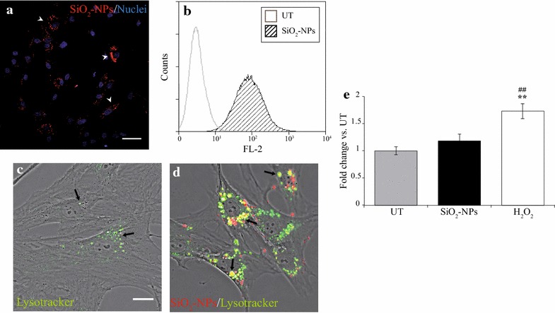
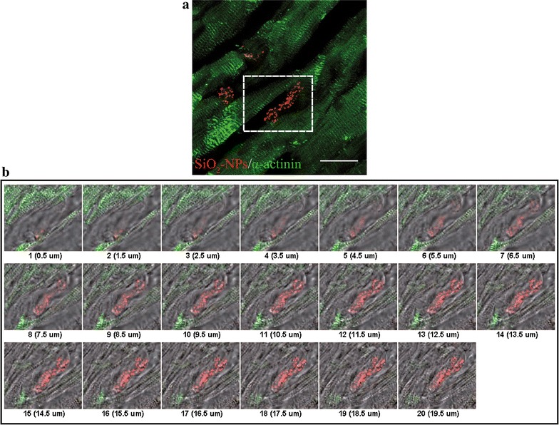
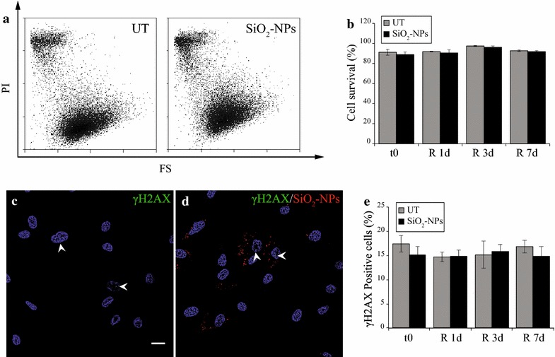
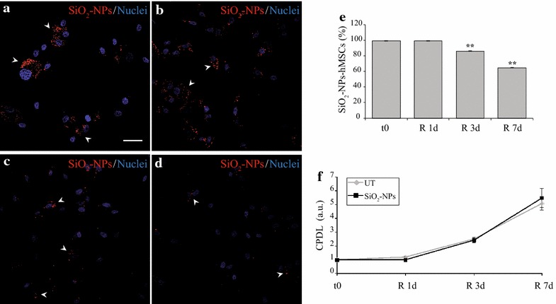
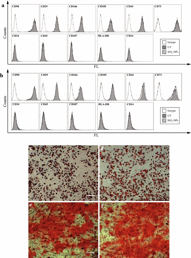
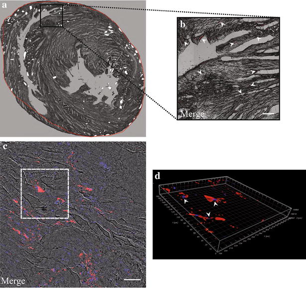
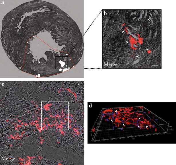
Optimal cell labelling is obtained after 16 h exposure of hMSCs to fluorescent 50 nm SiO2-NPs (50 µg mL(-1)); interestingly, lysosomal activation consequent to NPs storage is not associated to oxidative stress. During prolonged culture hMSCs do not undergo cyto- or genotoxicity, preserve their proliferative potential and their stemness/differentiation properties. Finally, the bright fluorescence emitted by internalized SiO2-NPs allows both clear visualization of hMSCs in normal and infarcted rat hearts and ultrastructural analysis of cell engraftment inside myocardial tissue.
Overall, 50 nm SiO2-NPs display elevated compatibility with hMSCs in terms of lack of cyto- and genotoxicity and maintenance of important features of these cells. The demonstrated biosafety, combined with proper cell labelling and visualization in histological sections, make these SiO2-NPs optimal candidates for the purpose of stem cell tracking inside heart tissue.
在动物和临床研究中,间充质干细胞(MSCs)治疗心肌梗死已被证明具有有益效果。工程化二氧化硅纳米颗粒(SiO2-NPs)因其抗降解性和易于功能化,已被广泛用作再生医学中的造影剂。然而,它们在细胞系统上的有效生物安全性仍存在争议。从这个角度来看,本研究的目的是:1)深入研究无定形50 nm SiO2-NPs对人骨髓来源的间充质干细胞(hMSCs)活力和功能的影响;2)优化hMSCs无害标记方案,并在跳动心脏模型中测试其可行性。
hMSCs暴露于荧光50 nm SiO2-NPs(50 µg mL(-1))16小时后可获得最佳细胞标记;有趣的是,纳米颗粒储存导致的溶酶体激活与氧化应激无关。在长期培养过程中,hMSCs不会发生细胞毒性或基因毒性,保持其增殖潜力以及干性/分化特性。最后,内化的SiO2-NPs发出的明亮荧光使得在正常和梗死大鼠心脏中都能清晰观察到hMSCs,并对心肌组织内的细胞植入进行超微结构分析。
总体而言,50 nm SiO2-NPs在缺乏细胞毒性和基因毒性以及维持这些细胞的重要特征方面与hMSCs具有高度兼容性。所证明的生物安全性,结合在组织切片中适当的细胞标记和可视化,使这些SiO2-NPs成为心脏组织内干细胞追踪的最佳候选物。