Reproductive Medicine Center, 105th Hospital of PLA, Hefei, 230031, Anhui, People's Republic of China.
The First affiliated Hospital of Anhui Medical University, Hefei, 230032, Anhui, People's Republic of China.
Stem Cell Res Ther. 2019 Aug 14;10(1):250. doi: 10.1186/s13287-019-1327-5.
Premature ovarian insufficiency (POI) is one of the leading causes of female infertility, which is caused by an abnormal ovarian reserve. Currently, there is no effective treatment to restore the fertility of POI patients. Recent studies suggested that microvesicles (MVs) released from mesenchymal stem cells (MSCs) exert therapeutic effects in various degenerative diseases. In this study, the effect of human umbilical cord MSC-derived MVs (HUCMSC-MVs) on the restoration of ovarian function in a chemotherapy-induced POI mouse model is investigated.
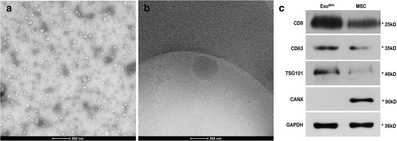
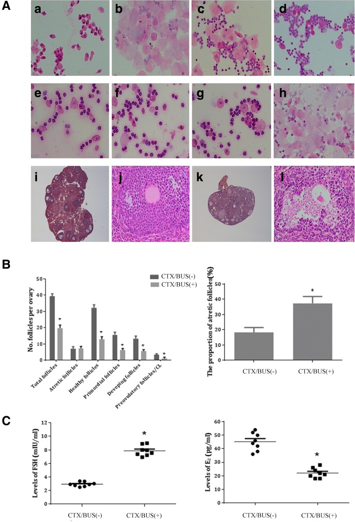
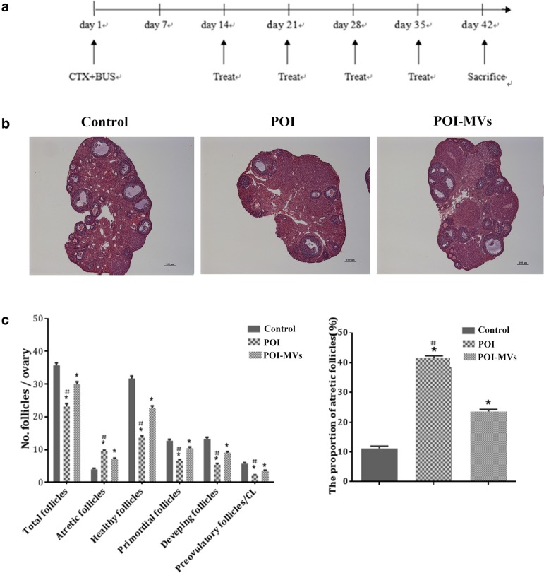
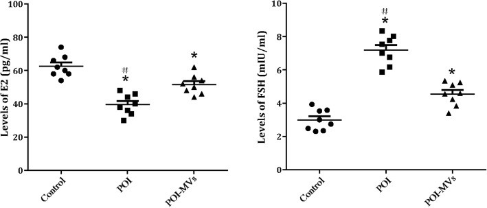
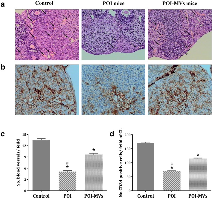
MVs were obtained from the supernatant of cultured HUCMSCs. The localization of PKH26-labeled HUCMSC-MVs in ovarian tissues was observed by confocal laser scanning microscopy. Histomorphometric analysis was performed to count the number of ovarian follicles and vessels. The ovarian sections were stained with anti-CD34 to evaluate angiogenesis. The levels of estradiol (E2) and follicle-stimulating hormone (FSH) were measured by enzyme-linked immunosorbent serologic assay. The mRNA expression of angiogenesis-related cytokines and the protein expression of AKT in mouse ovaries were measured by quantitative RT-PCR and western blot analysis. The parametric variables were compared by Student's t test and analysis of variance. The non-parametric variables were compared by the Mann-Whitney U test. Categorical variables were compared by χ test. P < 0.05 was considered statistically significant.
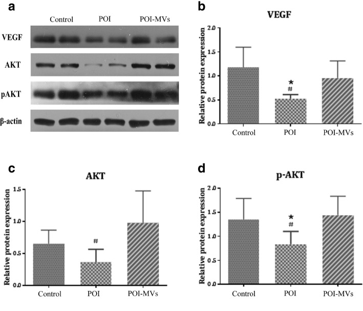
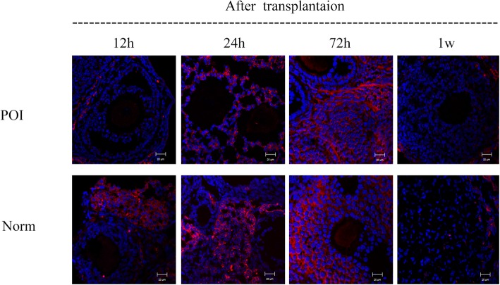
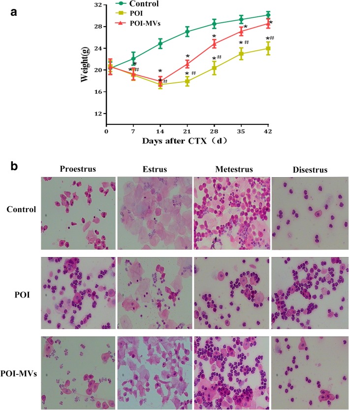
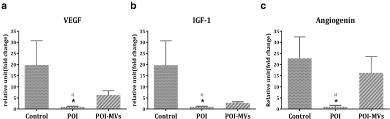
PKH26-labeled HUCMSC-MVs were detectable within the ovaries and migrated to the ovarian follicles 24 h after transplantation. The transplantation of HUCMSC-MVs could increase the body weight and number of ovarian follicles (primordial, developing, and preovulatory follicles), induce ovarian angiogenesis, and recover the disturbed estrous cycle of POI mice. The expression levels of total AKT, p-AKT, and angiogenic cytokines (including VEGF, IGF, and angiogenin) in the ovaries of POI mice were markedly upregulated after HUCMSC-MVs transplantation, suggesting that HUCMSC-MVs transplantation might recover ovarian function by inducing angiogenesis via the PI3K/AKT signaling pathway.
This study provides valuable insight into the effects of HUCMSC-MVs on ovarian tissue angiogenesis and on the restoration of ovarian function in POI mice, which may be helpful to develop a treatment strategy for POI patients.
卵巢早衰(POI)是女性不孕的主要原因之一,其病因是卵巢储备功能异常。目前,尚无有效的治疗方法可以恢复 POI 患者的生育能力。最近的研究表明,间充质干细胞(MSCs)释放的微小囊泡(MVs)在各种退行性疾病中具有治疗作用。在这项研究中,研究了人脐带 MSC 衍生的 MVs(HUCMSC-MVs)对化疗诱导的 POI 小鼠模型中卵巢功能恢复的影响。
从培养的 HUCMSCs 的上清液中获得 MVs。通过共聚焦激光扫描显微镜观察 PKH26 标记的 HUCMSC-MVs 在卵巢组织中的定位。进行组织形态计量学分析以计数卵巢卵泡和血管的数量。用抗 CD34 对卵巢切片进行染色以评估血管生成。通过酶联免疫吸附血清学测定法测量雌二醇(E2)和卵泡刺激素(FSH)的水平。通过定量 RT-PCR 和 Western blot 分析测量卵巢中血管生成相关细胞因子的 mRNA 表达和 AKT 的蛋白表达。通过学生 t 检验和方差分析比较参数变量。通过 Mann-Whitney U 检验比较非参数变量。通过 χ 检验比较分类变量。P<0.05 被认为具有统计学意义。
PKH26 标记的 HUCMSC-MVs 可在卵巢内检测到,并在移植后 24 小时迁移到卵巢卵泡中。HUCMSC-MVs 的移植可以增加 POI 小鼠的体重和卵巢卵泡数量(原始卵泡、发育卵泡和排卵前卵泡),诱导卵巢血管生成,并恢复 POI 小鼠紊乱的发情周期。POI 小鼠卵巢中总 AKT、p-AKT 和血管生成细胞因子(包括 VEGF、IGF 和血管生成素)的表达水平在 HUCMSC-MVs 移植后明显上调,提示 HUCMSC-MVs 移植可能通过 PI3K/AKT 信号通路诱导血管生成来恢复卵巢功能。
这项研究提供了关于 HUCMSC-MVs 对卵巢组织血管生成和 POI 小鼠卵巢功能恢复影响的有价值的见解,这可能有助于为 POI 患者开发治疗策略。