Tissue Engineering Centre, Faculty of Medicine, Universiti Kebangsaan Malaysia, Kuala Lumpur, Malaysia.
Department of Orthopedic & Traumatology, Faculty of Medicine, Universiti Kebangsaan Malaysia, Kuala Lumpur, Malaysia.
Indian J Med Res. 2019 May;149(5):641-649. doi: 10.4103/ijmr.IJMR_45_17.
BACKGROUND & OBJECTIVES: Seeding density is one of the major parameters affecting the quality of tissue-engineered cartilage. The objective of this study was to evaluate different seeding densities of osteoarthritis chondrocytes (OACs) to obtain the highest quality cartilage.
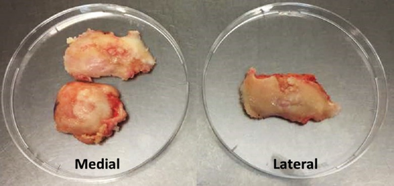
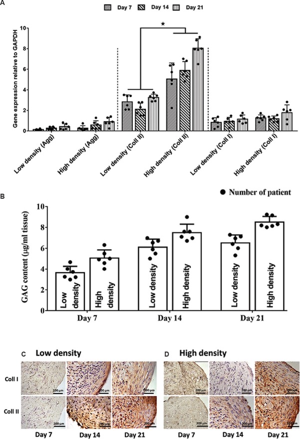
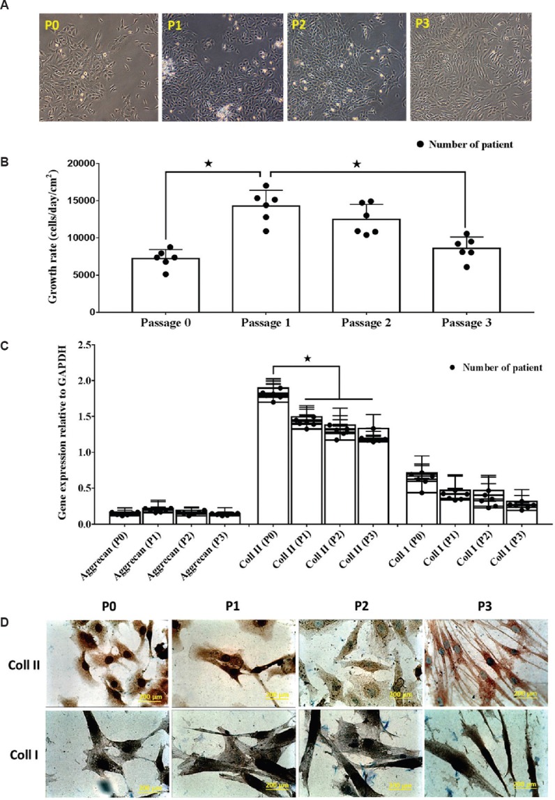
The OACs were expanded from passage 0 (P0) to P3, and cells in each passage were analyzed for gross morphology, growth rate, RNA expression and immunochemistry (IHC). The harvested OACs were assigned into two groups: low (1×10 cells/ml) and high (3×10 cells/ml) cell density. Three-dimensional (3D) constructs for each group were created using polymerised fibrin and cultured for 7, 14 and 21 days in vitro using chondrocyte growth medium. OAC constructs were analyzed with gross assessments and microscopic evaluation using standard histology, IHC and immunofluorescence staining, in addition to gene expression and biochemical analyses to evaluate tissue development.
Constructs with a high seeding density of 3×10 cells/ml were associated with better quality cartilage-like tissue than those seeded with 1×10 cells/ml based on overall tissue formation, cell association and extracellular matrix distribution. The chondrogenic properties of the constructs were further confirmed by the expression of genes encoding aggrecan core protein and collagen type II.
INTERPRETATION & CONCLUSIONS: Our results confirmed that cell density was a significant factor affecting cell behaviour and aggregate production, and this was important for establishing good quality cartilage.
接种密度是影响组织工程软骨质量的主要参数之一。本研究旨在评估不同的骨关节炎软骨细胞(OAC)接种密度,以获得最佳质量的软骨。
将 OAC 从第 0 代(P0)扩增至第 3 代(P3),并对每一代的细胞进行大体形态、生长速度、RNA 表达和免疫化学(IHC)分析。收获的 OAC 被分为两组:低(1×10 个细胞/ml)和高(3×10 个细胞/ml)细胞密度。使用聚合纤维蛋白为每组创建三维(3D)构建体,并在体外使用软骨细胞生长培养基分别培养 7、14 和 21 天。通过大体评估和标准组织学、IHC 和免疫荧光染色的微观评估,以及基因表达和生化分析来评估组织发育,对 OAC 构建体进行分析。
基于总体组织形成、细胞关联和细胞外基质分布,以 3×10 个细胞/ml 的高接种密度构建的软骨样组织比以 1×10 个细胞/ml 的接种密度构建的软骨样组织具有更好的质量。构建体的软骨形成特性进一步通过编码聚集蛋白核心蛋白和 II 型胶原的基因的表达得到证实。
我们的结果证实细胞密度是影响细胞行为和聚集产物的重要因素,这对于建立高质量的软骨很重要。