Department of Surgery, Duke University, Durham, NC, United States.
Front Immunol. 2019 Jul 31;10:1767. doi: 10.3389/fimmu.2019.01767. eCollection 2019.
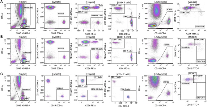
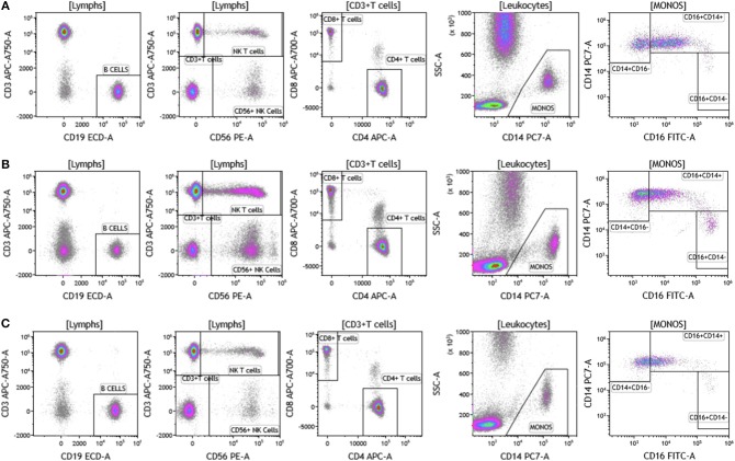
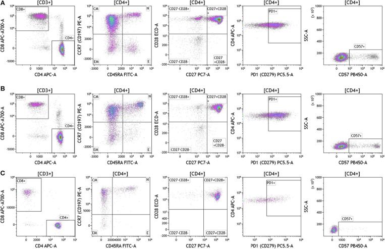
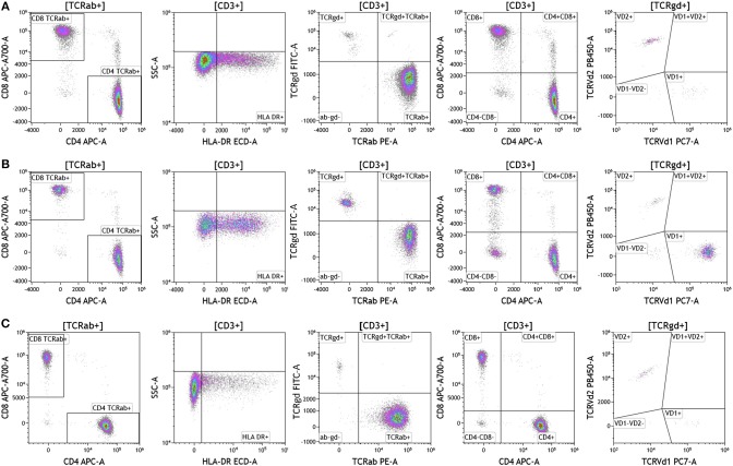
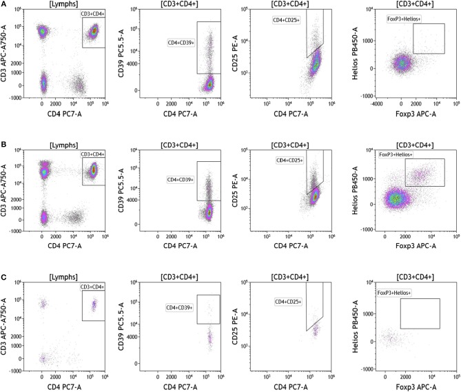
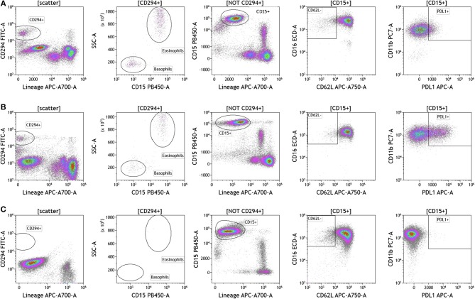
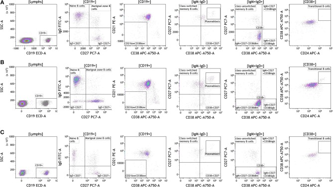
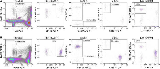
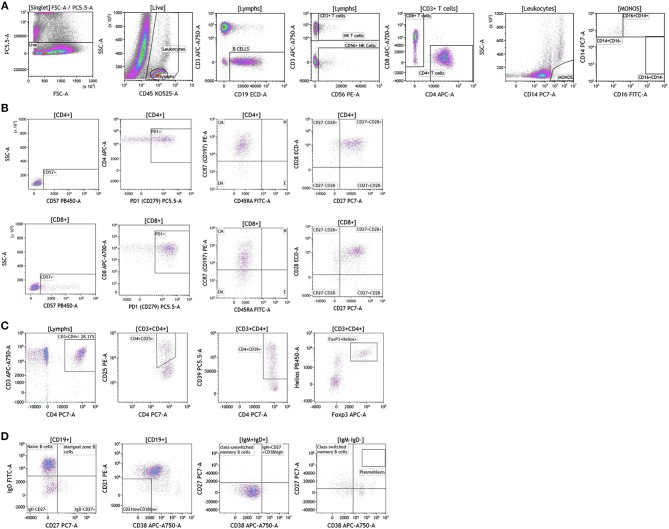
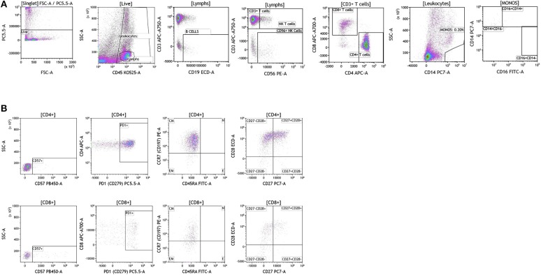
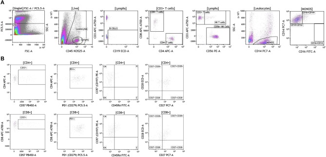
Immunotherapies are rapidly being integrated into standard of care (SOC) therapy in conjunction with surgery, chemotherapy, and radiotherapy for many cancers and a large number of clinical studies continue to explore immunotherapy alone and as part of combination therapies in patients with cancer. It is evident that clinical effectiveness of immunotherapy is limited to a subset of patients and improving immunotherapy related outcomes remains a major scientific and clinical effort. Understanding the immune cell subset phenotype and activation/functional status (cellular immunome) prior to and post therapy is therefore critical to develop biomarkers that (1) will predict if a patient will respond to immunotherapy and (2) are a result of immunotherapy. In this study, we investigated local (tumor) and peripheral (blood) cellular immunome of patients with melanoma, breast cancer, and brain cancer using a rapid and reliable standardized, multiparameter flow cytometry assay. We used this approach to monitor changes in the peripheral cellular immunome in women with breast cancer undergoing SOC therapy. Our analysis is unique because it is conducted using matched fresh tumor tissue and blood from patients in real-time, within 2-3 h of sample acquisition, and provides insight into the innate and adaptive immune cell profile in blood and tumor. Specific to blood, this approach involves no manipulation and evaluates all immune subsets such as T cells, B cells, natural killer (NK) cells, monocytes, dendritic cells (DCs), neutrophils, eosinophils, and basophils using 0.5 ml of blood. Analysis of the corresponding tumor provides much needed insight into the phenotype and activation status of immune cells, especially T and B cells, in the tumor microenvironment vs. the periphery. This analysis will be used to assess baseline and therapy-mediated changes in local and peripheral cellular immunome in patients with glioblastoma, breast cancer, and melanoma in planned immunotherapy clinical studies.
免疫疗法正在与手术、化疗和放疗相结合,迅速被纳入许多癌症的标准治疗(SOC)方案中,大量的临床研究继续探索免疫疗法单独应用以及作为癌症患者联合治疗的一部分。显然,免疫疗法的临床效果仅限于一部分患者,提高免疫疗法相关的疗效仍然是一项重大的科学和临床努力。因此,在治疗前后了解免疫细胞亚群表型和激活/功能状态(细胞免疫组)对于开发预测患者是否对免疫疗法有反应的生物标志物以及评估免疫疗法的结果至关重要。在这项研究中,我们使用一种快速可靠的标准化多参数流式细胞术检测方法,研究了黑色素瘤、乳腺癌和脑癌患者的局部(肿瘤)和外周(血液)细胞免疫组。我们使用这种方法来监测接受 SOC 治疗的乳腺癌女性外周细胞免疫组的变化。我们的分析是独特的,因为它是使用实时从患者获得的新鲜肿瘤组织和血液样本进行的,在样本采集后 2-3 小时内完成,可以深入了解血液和肿瘤中的固有和适应性免疫细胞谱。具体到血液,这种方法无需操作,可评估所有免疫亚群,如 T 细胞、B 细胞、自然杀伤(NK)细胞、单核细胞、树突状细胞(DC)、中性粒细胞、嗜酸性粒细胞和嗜碱性粒细胞,仅需 0.5 毫升血液。对相应肿瘤的分析提供了对肿瘤微环境中与外周免疫细胞的表型和激活状态的深入了解,特别是 T 和 B 细胞。这项分析将用于评估计划中的胶质母细胞瘤、乳腺癌和黑色素瘤免疫治疗临床研究中患者的局部和外周细胞免疫组的基线和治疗介导的变化。