Department of Pharmacology and Dental Therapeutics, Program in Neurobiology, Dental Research Institute, Seoul National University School of Dentistry, Seoul, Republic of Korea.
Department of Dental Anesthesiology, Program in Neurobiology, Dental Research Institute, Seoul National University School of Dentistry, Seoul, Republic of Korea.
PLoS One. 2019 Aug 16;14(8):e0221156. doi: 10.1371/journal.pone.0221156. eCollection 2019.
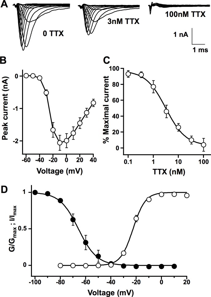
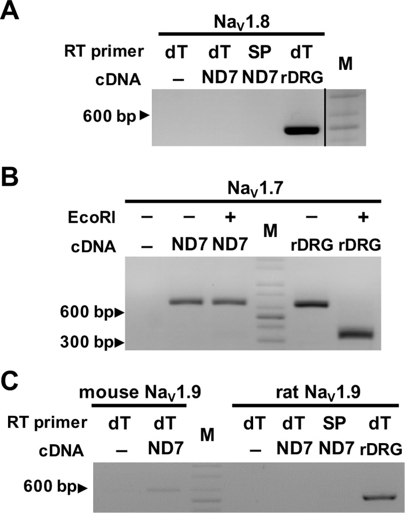
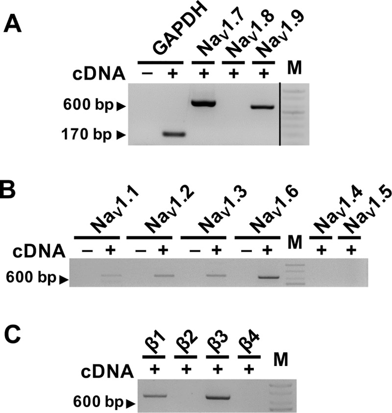
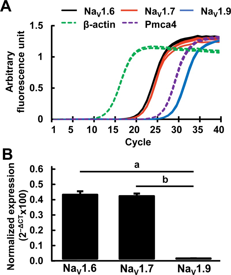
ND7/23 cells are gaining traction as a host model to express peripheral sodium channels such as NaV1.8 and NaV1.9 that have been difficult to express in widely utilized heterologous cells, like CHO and HEK293. Use of ND7/23 as a model cell to characterize the properties of sodium channels requires clear understanding of the endogenous ion channels. To define the nature of the background sodium currents in ND7/23 cells, we aimed to comprehensively profile the voltage-gated sodium channel subunits by endpoint and quantitative reverse transcription-PCR and by whole-cell patch clamp electrophysiology. We found that untransfected ND7/23 cells express endogenous peak sodium currents that average -2.12nA (n = 15) and with kinetics typical of fast sodium currents having activation and inactivation completed within few milliseconds. Furthermore, sodium currents were reduced to virtually nil upon exposure to 100nM tetrodotoxin, indicating that ND7/23 cells have essentially null background for tetrodotoxin-resistant (TTX-R) currents. qRT-PCR profiling indicated a major expression of TTX-sensitive (TTX-S) NaV1.6 and NaV1.7 at similar levels and very low expression of TTX-R NaV1.9 transcripts. There was no expression of TTX-R NaV1.8 in ND7/23 cells. There was low expression of NaV1.1, NaV1.2, NaV1.3 and no expression of cardiac or skeletal muscle sodium channels. As for the sodium channel auxiliary subunits, β1 and β3 subunits were expressed, but not the β2 and β4 subunits that covalently associate with the α-subunits. In addition, our results also showed that only the mouse forms of NaV1.6, NaV1.7 and NaV1.9 sodium channels were expressed in ND7/23 cells that was originally generated as a hybridoma of rat embryonic DRG and mouse neuroblastoma cell-line. By molecular profiling of auxiliary β- and principal α-subunits of the voltage gated sodium channel complex, our results define the background sodium channels expressed in ND7/23 cells, and confirm their utility for detailed functional studies of emerging pain channelopathies ascribed to mutations of the TTX-R sodium channels of sensory neurons.
ND7/23 细胞作为一种表达外周钠通道(如 NaV1.8 和 NaV1.9)的宿主模型而备受关注,这些钠通道在广泛应用的异源细胞(如 CHO 和 HEK293)中很难表达。使用 ND7/23 作为模型细胞来表征钠通道的特性,需要清楚地了解内源性离子通道。为了全面描绘 ND7/23 细胞中电压门控钠通道亚基的特性,我们旨在通过终点和定量逆转录 PCR 以及全细胞膜片钳电生理学来全面描绘电压门控钠通道亚基。我们发现,未转染的 ND7/23 细胞表达内源性峰值钠电流,平均为-2.12nA(n=15),其动力学特征为快速钠电流,激活和失活在几毫秒内完成。此外,暴露于 100nM 河豚毒素后,钠电流几乎降至零,表明 ND7/23 细胞的河豚毒素抗性(TTX-R)电流背景基本为零。qRT-PCR 分析表明,TTX-S NaV1.6 和 NaV1.7 的主要表达水平相似,TTX-R NaV1.9 转录本的表达水平非常低。ND7/23 细胞中没有 TTX-R NaV1.8 的表达。NaV1.1、NaV1.2 和 NaV1.3 的表达水平较低,没有表达心肌或骨骼肌钠通道。至于钠通道辅助亚基,β1 和 β3 亚基表达,但不表达与 α-亚基共价结合的 β2 和 β4 亚基。此外,我们的结果还表明,只有 NaV1.6、NaV1.7 和 NaV1.9 钠通道的鼠形式在 ND7/23 细胞中表达,该细胞最初是由大鼠胚胎背根神经节和小鼠神经母细胞瘤细胞系的杂交瘤产生的。通过对电压门控钠通道复合物的辅助β-和主要α-亚基的分子分析,我们的结果定义了 ND7/23 细胞中表达的背景钠通道,并证实了它们在新兴疼痛通道病的详细功能研究中的效用,这些通道病归因于感觉神经元 TTX-R 钠通道的突变。