Research and Development, Organogenesis, 2641 Rock Ridge Lane Birmingham, Alabama, 35216.
Wound Repair Regen. 2019 Nov;27(6):609-621. doi: 10.1111/wrr.12757. Epub 2019 Aug 29.
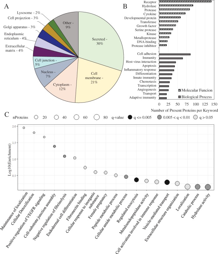
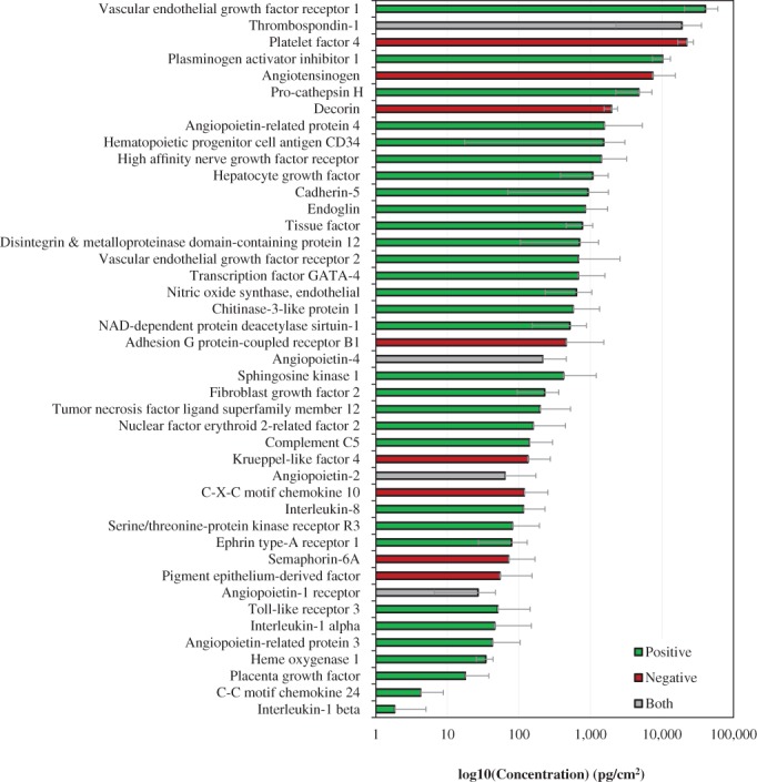
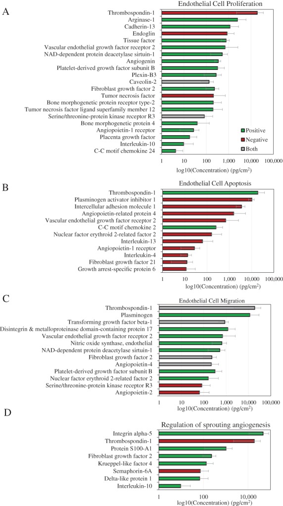
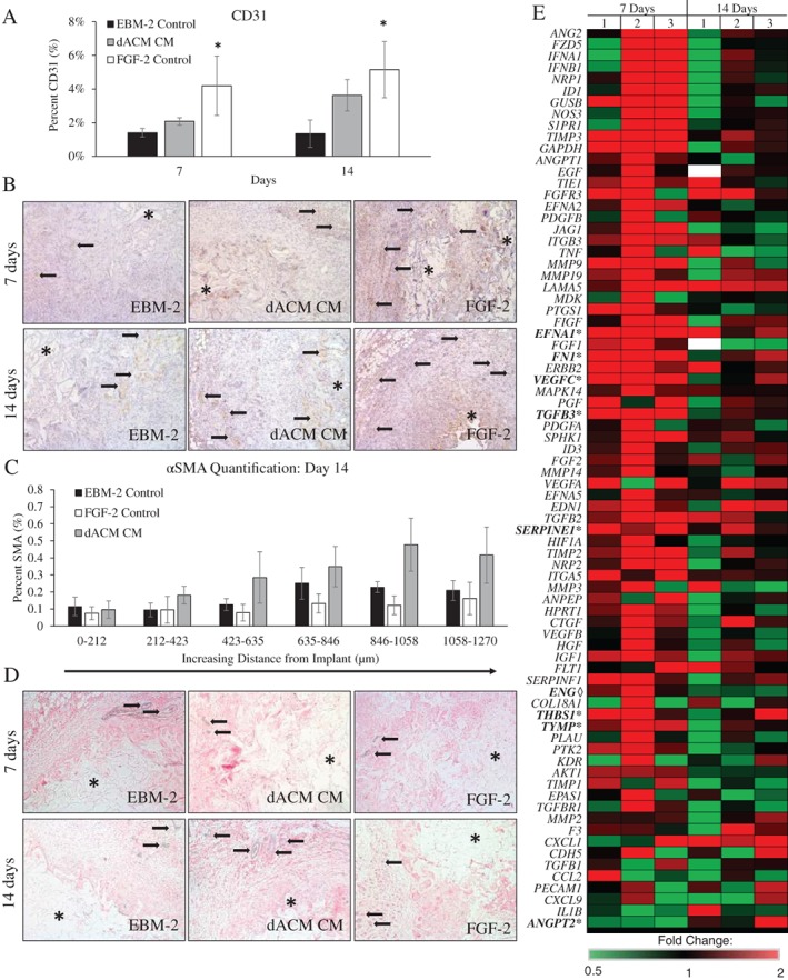
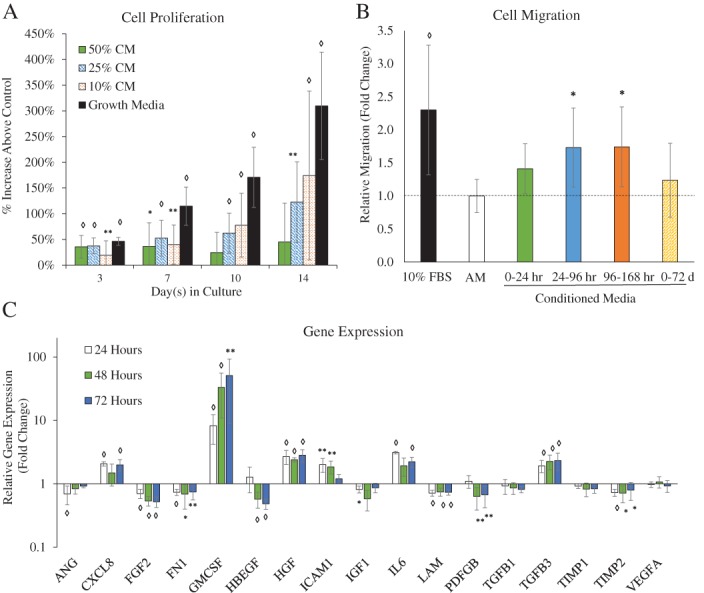
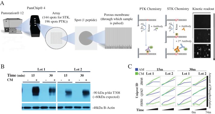
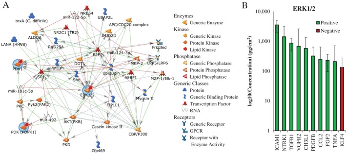
Angiogenesis is essential for the successful repair of tissues; however, in many chronic conditions, angiogenesis is inhibited. Placental tissues have been shown to illicit an angiogenic response both in vitro and in vivo, and the angiogenic properties of these tissues likely contribute to observed clinical outcomes. Although there is some work describing the angiogenic effects of these tissues, comparatively little has been done to determine the possible mechanisms responsible for this effect. The purpose of this study was to conduct a thorough evaluation of a commercially available dehydrated amnion chorion membrane to better understand how these tissues may promote angiogenesis. The proteomic content of this tissue was evaluated using a high throughput proteomic microarray, and then the effects of these grafts were evaluated in vivo using subcutaneous gelfoam sponge implants containing conditioned media (CM) from the graft. Human microvascular endothelial cells were then used to determine how released factors effect migration, proliferation, gene expression, and protein production in vitro. Finally, to elucidate potential signaling-pathways through which tissue-derived factors act to induce pro-angiogenetic phenotypes in endothelial cells in vitro, we performed a global analysis of both serine/threonine and tyrosine kinase activity. Kinomic and proteomic data were then combined to generate protein-protein interaction networks that enabled the identification of multiple growth factors and cytokines with both pro- and anti-angiogenetic properties. In vivo, the addition of CM resulted in increased CD31 and αSMA staining and increases in pro-angiogenic gene expression. In vitro, CM resulted in significant increases in endothelial proliferation, migration, and the expression of granulocyte-macrophage colony-stimulating factor, hepatocyte growth factor, and transforming growth factor beta-3. Integrated kinomic analysis implicated ERK1/2 signaling as the primary pathway activated following culture of endothelial cells with dehydrated amnion/chorion membrane (dACM) CM. In conclusion, dACM grafts triggered pro-angiogenic responses both in vitro and in vivo that are likely at least partially mediated by ERK1/2 signaling.
血管生成对于组织的成功修复至关重要;然而,在许多慢性疾病中,血管生成受到抑制。胎盘组织已被证明在体外和体内均能引起血管生成反应,这些组织的血管生成特性可能有助于观察到的临床结果。虽然有一些工作描述了这些组织的血管生成作用,但相对较少的工作是确定负责这种作用的可能机制。本研究的目的是对一种商业上可获得的脱水羊膜绒毛膜膜进行全面评估,以更好地了解这些组织如何促进血管生成。使用高通量蛋白质组微阵列评估了该组织的蛋白质组内容,然后通过含有来自移植物的条件培养基 (CM) 的皮下明胶海绵植入物在体内评估这些移植物的效果。然后,使用人微血管内皮细胞来确定释放的因子如何在体外影响迁移、增殖、基因表达和蛋白质产生。最后,为了阐明组织衍生因子在体外诱导内皮细胞形成促血管生成表型的潜在信号通路,我们对丝氨酸/苏氨酸和酪氨酸激酶活性进行了全局分析。激酶组学和蛋白质组学数据随后被组合在一起,生成蛋白质-蛋白质相互作用网络,从而鉴定出具有促血管生成和抗血管生成特性的多种生长因子和细胞因子。在体内,CM 的添加导致 CD31 和αSMA 染色增加,并增加了促血管生成基因的表达。在体外,CM 导致内皮细胞增殖、迁移以及粒细胞-巨噬细胞集落刺激因子、肝细胞生长因子和转化生长因子β-3 的表达显著增加。综合激酶组学分析表明,在培养内皮细胞时,ERK1/2 信号通路被激活,随后 ERK1/2 信号通路被激活,这可能至少部分是由脱水羊膜/绒毛膜膜 (dACM) CM 介导的。总之,dACM 移植物在体外和体内均引发促血管生成反应,这些反应可能至少部分是由 ERK1/2 信号通路介导的。