School of Biomedical Engineering, Korea University, Seoul, South Korea.
School of Engineering, Brown University, Providence, Rhode Island, United States of America.
PLoS One. 2019 Aug 22;14(8):e0220810. doi: 10.1371/journal.pone.0220810. eCollection 2019.
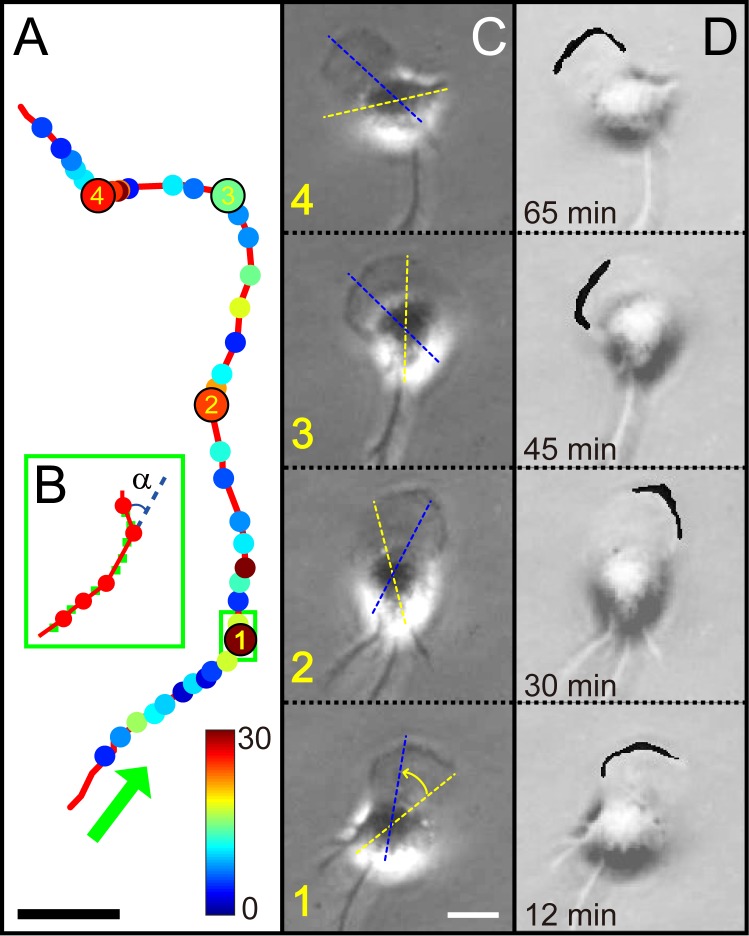
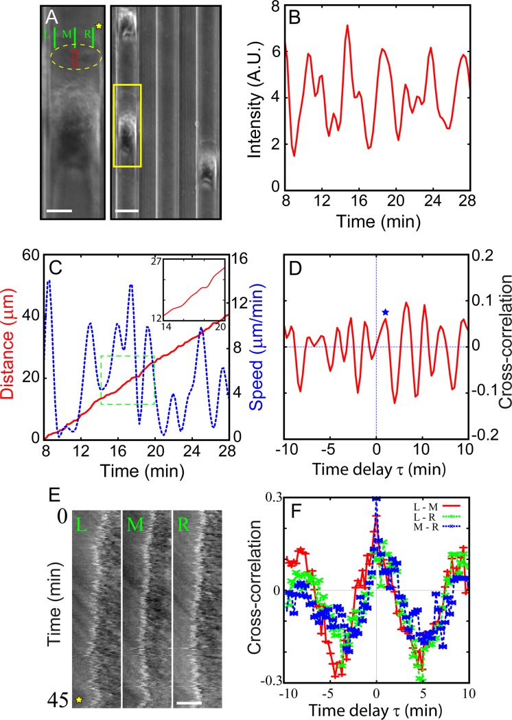
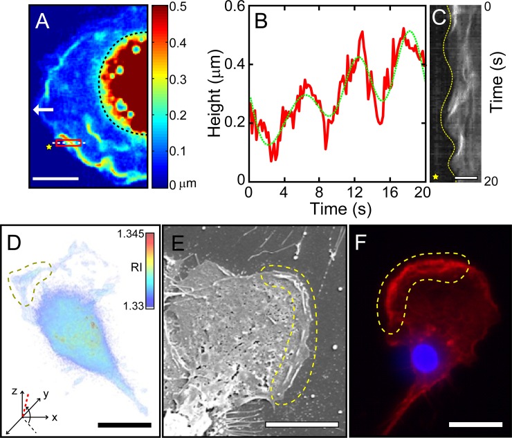
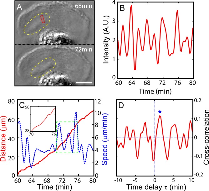
Freely crawling cells are often viewed as randomly moving Brownian particles but they generally exhibit some directional persistence. This property is often related to their zigzag motile behaviors that can be described as a noisy but temporally structured sequence of "runs" and "turns." However, its underlying biophysical mechanism is largely unexplored. Here, we carefully investigate the collective actin wave dynamics associated with the zigzag-crawling movements of microglia (as primary brain immune cells) employing a number of different quantitative imaging modalities including synthetic aperture microscopy and optical diffraction tomography, as well as conventional fluorescence imaging and scanning electron microscopy. Interestingly, we find that microglia exhibit two distinct types of actin waves working at two quite different time scales and locations, and they seem to serve different purposes. One type of actin waves is fast "peripheral ruffles" arising spontaneously with an oscillating period of about 6 seconds at some portion of the leading edge of crawling microglia, where the vigorously biased peripheral ruffles seem to set the direction of a new turn (in 2-D free space). When the cell turning events are inhibited with a physical confinement (in 1-D track), the peripheral ruffles still exist at the leading edge with no bias but showing phase coherence in the cell crawling direction. The other type is "dorsal actin waves" which also exhibits an oscillatory behavior but with a much longer period of around 2 minutes compared to the fast "peripheral ruffles". Dorsal actin waves (whether the cell turning events are inhibited or not) initiate in the lamellipodium just behind the leading edge, travelling down toward the core region of the cell and disappear. Such dorsal wave propagations seem to be correlated with migration of the cell. Thus, we may view the dorsal actin waves are connected with the "run" stage of cell body, whereas the fast ruffles at the leading front are involved in the "turn" stage.
自由爬行的细胞通常被视为随机移动的布朗粒子,但它们通常表现出一定的方向持续性。这种性质通常与它们的之字形运动行为有关,可以描述为一个嘈杂但具有时间结构的“奔跑”和“转弯”序列。然而,其潜在的生物物理机制在很大程度上尚未被探索。在这里,我们使用多种不同的定量成像模式,包括合成孔径显微镜和光学衍射断层扫描,以及传统的荧光成像和扫描电子显微镜,仔细研究了与小胶质细胞(作为主要的大脑免疫细胞)之字形爬行运动相关的集体肌动蛋白波动力学。有趣的是,我们发现小胶质细胞表现出两种不同类型的肌动蛋白波,它们在两个非常不同的时间尺度和位置上工作,并且它们似乎服务于不同的目的。一种类型的肌动蛋白波是快速的“周边隆起”,自发地在爬行小胶质细胞的前缘的一部分以约 6 秒的振荡周期出现,在那里,剧烈偏向的周边隆起似乎设定了新转弯的方向(在二维自由空间中)。当用物理限制(在一维轨迹中)抑制细胞转弯事件时,前缘仍然存在周边隆起,但没有偏向性,而是在细胞爬行方向上表现出相位相干性。另一种是“背侧肌动蛋白波”,它也表现出振荡行为,但与快速的“周边隆起”相比,其周期要长得多,约为 2 分钟。背侧肌动蛋白波(无论是否抑制细胞转弯事件)在前缘后面的片状伪足中起始,向下传播到细胞的核心区域并消失。这种背向波传播似乎与细胞的迁移有关。因此,我们可以将背侧肌动蛋白波与细胞体的“奔跑”阶段联系起来,而前缘的快速隆起则与“转弯”阶段有关。