García-Gomar María G, Strong Christian, Toschi Nicola, Singh Kavita, Rosen Bruce R, Wald Lawrence L, Bianciardi Marta
Department of Radiology, Athinoula A. Martinos Center for Biomedical Imaging, MGH and Harvard Medical School, Boston, MA, United States.
Department of Neurosurgery, Brigham and Women's Hospital, Harvard Medical School, Boston, MA, United States.
Front Neurosci. 2019 Aug 7;13:764. doi: 10.3389/fnins.2019.00764. eCollection 2019.
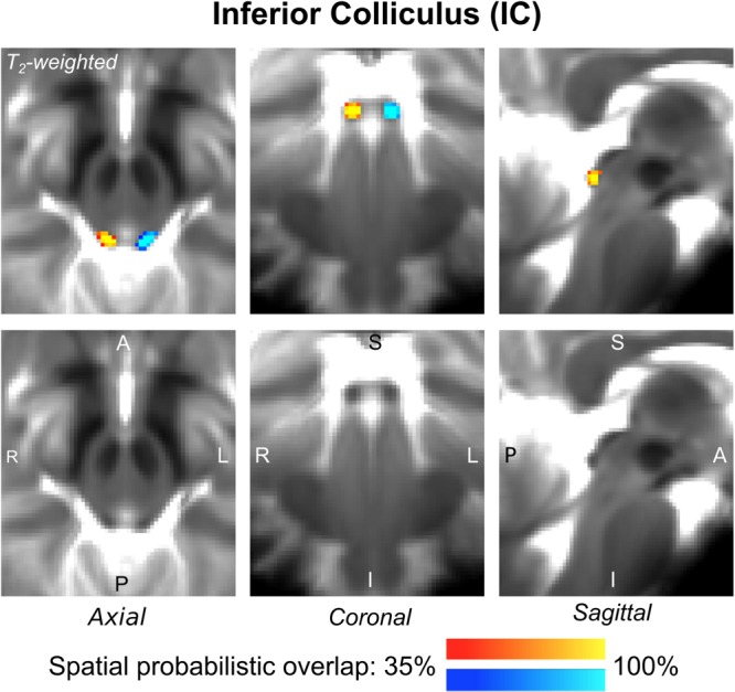
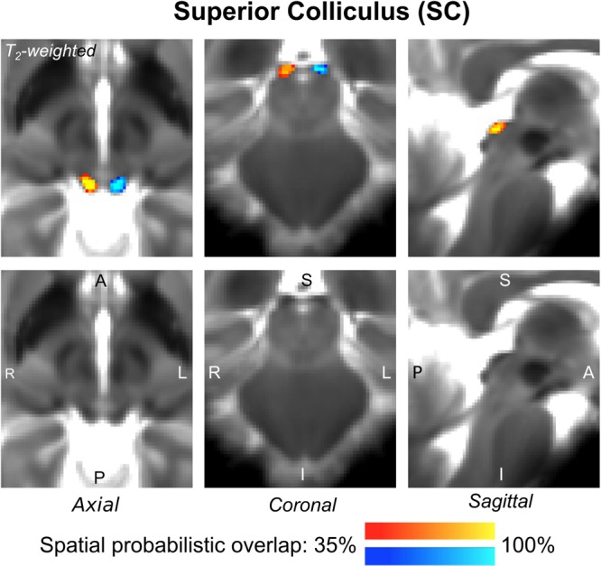
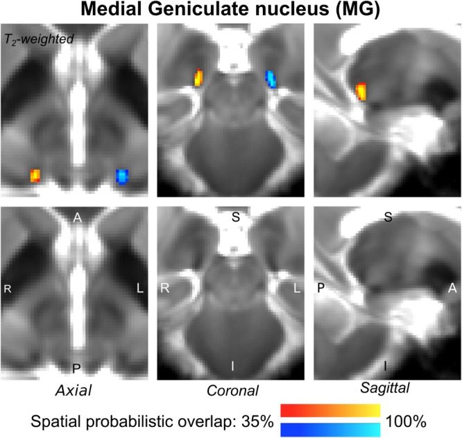
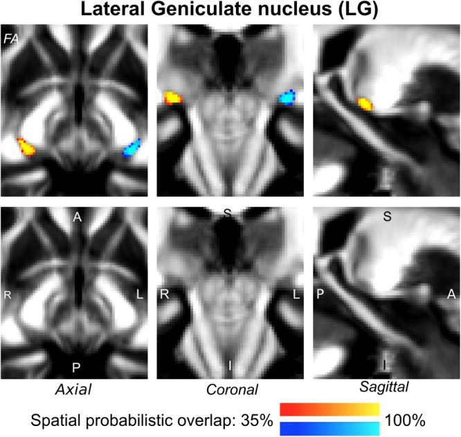
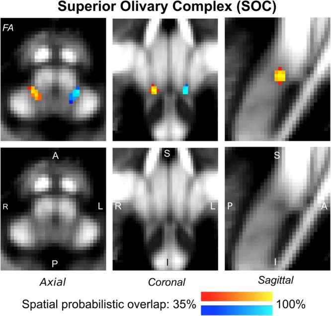
Despite extensive neuroimaging research of primary sensory cortices involved in auditory and visual functions, subcortical structures within these domains, such as the inferior and superior colliculi, the medial and lateral geniculate nuclei and the superior olivary complex, are currently understudied with magnetic resonance imaging (MRI) in living humans. This is because a precise localization of these nuclei is hampered by the limited contrast and sensitivity of conventional neuroimaging methods for deep brain nuclei. In this work, we used 7 Tesla multi-modal (T-weighted and diffusion fractional anisotropy) 1.1 mm isotropic resolution MRI to achieve high sensitivity and contrast for single-subject brainstem and thalamic nuclei delineation. After precise coregistration to stereotactic space, we generated an human probabilistic atlas of auditory (medial geniculate nucleus, inferior colliculus, and superior olivary complex) and visual (lateral geniculate nucleus and superior colliculus) subcortical nuclei. We foresee the use of this atlas as a tool to precisely identify the location and shape of auditory/visual deep nuclei in research as well as clinical human studies.
尽管对参与听觉和视觉功能的初级感觉皮层进行了广泛的神经影像学研究,但目前在活体人类中,利用磁共振成像(MRI)对这些区域内的皮层下结构,如下丘和上丘、内侧和外侧膝状体核以及上橄榄复合体的研究仍不足。这是因为传统神经成像方法对深部脑核的对比度和灵敏度有限,阻碍了这些核的精确定位。在这项研究中,我们使用7特斯拉多模态(T加权和扩散分数各向异性)1.1毫米各向同性分辨率MRI,以实现对单个体脑干和丘脑核的高灵敏度和对比度描绘。在精确配准到立体定向空间后,我们生成了一个人类听觉(内侧膝状体核、下丘和上橄榄复合体)和视觉(外侧膝状体核和上丘)皮层下核的概率图谱。我们预计将该图谱用作一种工具,以便在研究以及临床人体研究中精确识别听觉/视觉深部核的位置和形状。