Singh Kavita, Indovina Iole, Augustinack Jean C, Nestor Kimberly, García-Gomar María G, Staab Jeffrey P, Bianciardi Marta
Brainstem Imaging Laboratory, Department of Radiology, Athinoula A. Martinos Center for Biomedical Imaging, Massachusetts General Hospital and Harvard Medical School, Boston, MA, United States.
Department of Medicine and Surgery, Saint Camillus International University of Health and Medical Sciences, Rome, Italy.
Front Neurosci. 2020 Jan 23;13:1425. doi: 10.3389/fnins.2019.01425. eCollection 2019.
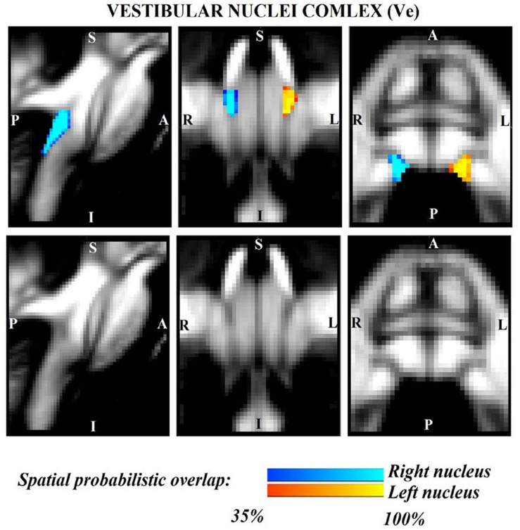
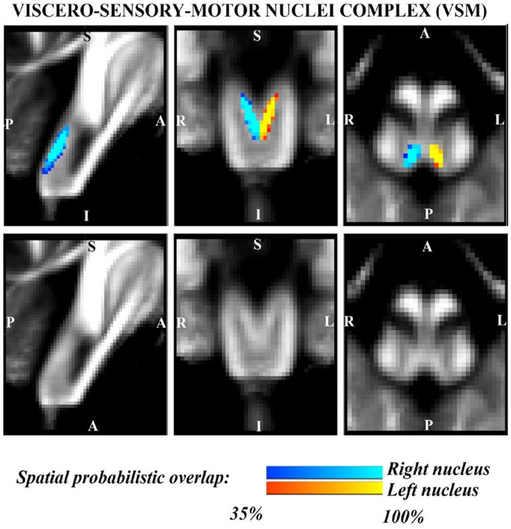
The lateral parabrachial nucleus, medial parabrachial nucleus, vestibular nuclei complex, and medullary viscero-sensory-motor (VSM) nuclei complex (the latter including among others the solitary nucleus, vagus nerve nucleus, and hypoglossal nucleus) are anatomically and functionally connected brainstem gray matter structures that convey signals across multiple modalities between the brain and the spinal cord to regulate vital bodily functions. It is remarkably difficult to precisely extrapolate the location of these nuclei from atlases to conventional 3 Tesla images; thus, a probabilistic brainstem template in stereotaxic neuroimaging space in living humans is needed. We delineated these nuclei using single-subject high contrast 1.1 mm isotropic resolution 7 Tesla MRI images. After precise coregistration of nuclei labels to stereotaxic space, we generated a probabilistic template of their anatomical locations. Finally, we validated the nuclei labels in the template by assessing their inter-rater agreement, consistency across subjects and volumes. We also performed a preliminary comparison of their location and microstructural properties to histologic sections of a postmortem human brainstem specimen. In future, the resulting probabilistic template of these brainstem nuclei in stereotaxic space may assist researchers and clinicians in evaluating autonomic, vestibular and VSM nuclei structure, function and connectivity in living humans using conventional 3 Tesla MRI scanners.
外侧臂旁核、内侧臂旁核、前庭神经核复合体以及延髓内脏感觉运动(VSM)核复合体(后者包括孤束核、迷走神经核和舌下神经核等)是在解剖学和功能上相连接的脑干灰质结构,它们在大脑和脊髓之间传递多种形式的信号,以调节身体的重要功能。要从图谱中精确推断出这些核在传统3特斯拉图像上的位置非常困难;因此,需要一个活体人类立体定向神经成像空间中的概率性脑干模板。我们使用单受试者高对比度1.1毫米各向同性分辨率的7特斯拉MRI图像描绘了这些核。在将核标签精确配准到立体定向空间后,我们生成了它们解剖位置的概率性模板。最后,我们通过评估评分者间的一致性、受试者间的一致性和体积,验证了模板中的核标签。我们还对它们的位置和微观结构特性与死后人类脑干标本的组织学切片进行了初步比较。未来,在立体定向空间中生成的这些脑干核的概率性模板可能会帮助研究人员和临床医生使用传统3特斯拉MRI扫描仪评估活体人类的自主神经、前庭和VSM核的结构、功能和连通性。