Wu Yi-Chia, Wang Ya-Chin, Wang Wei-Ting, Wang Hui-Min David, Lin Hsin-Hung, Su Long-Jyun, Kuo Yur-Ren, Lai Chung-Sheng, Ho Mei-Ling, Yu John
Institute of Stem Cell and Translational Cancer Research, Chang Gung Memorial Hospital at Linko, Taoyuan 333, Taiwan.
Ph.D. Program in Translational Medicine, Kaohsiung Medical University, Kaohsiung, and Academia Sinica, Taipei 115, Taiwan.
Polymers (Basel). 2019 Aug 23;11(9):1391. doi: 10.3390/polym11091391.
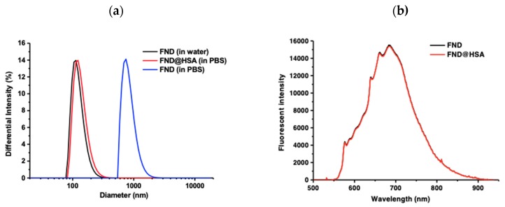
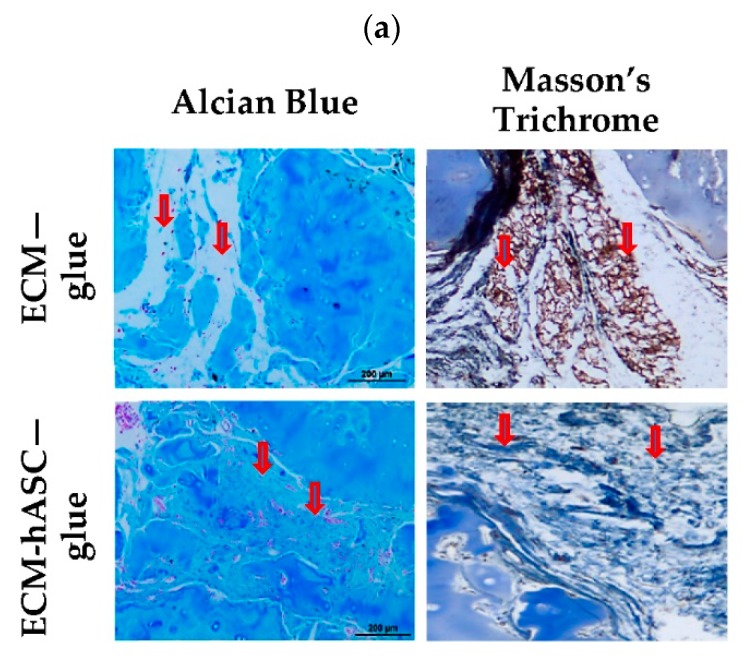
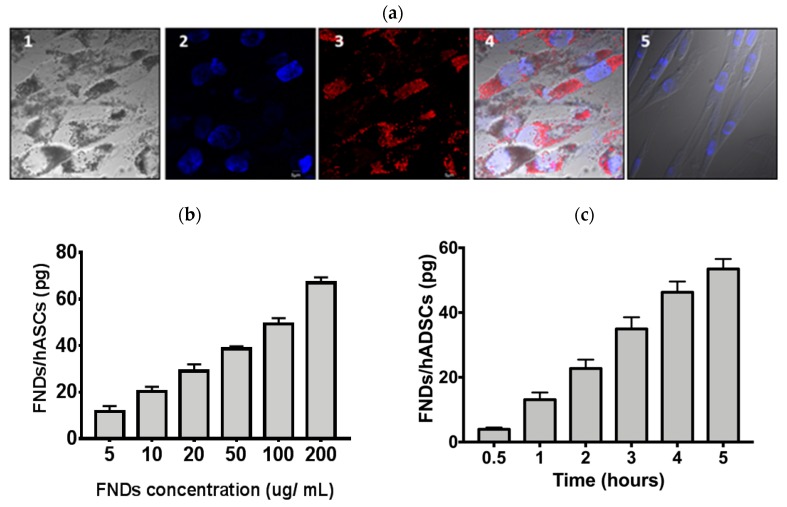
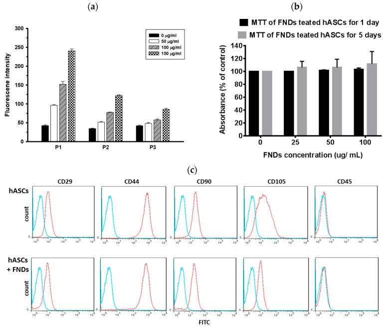
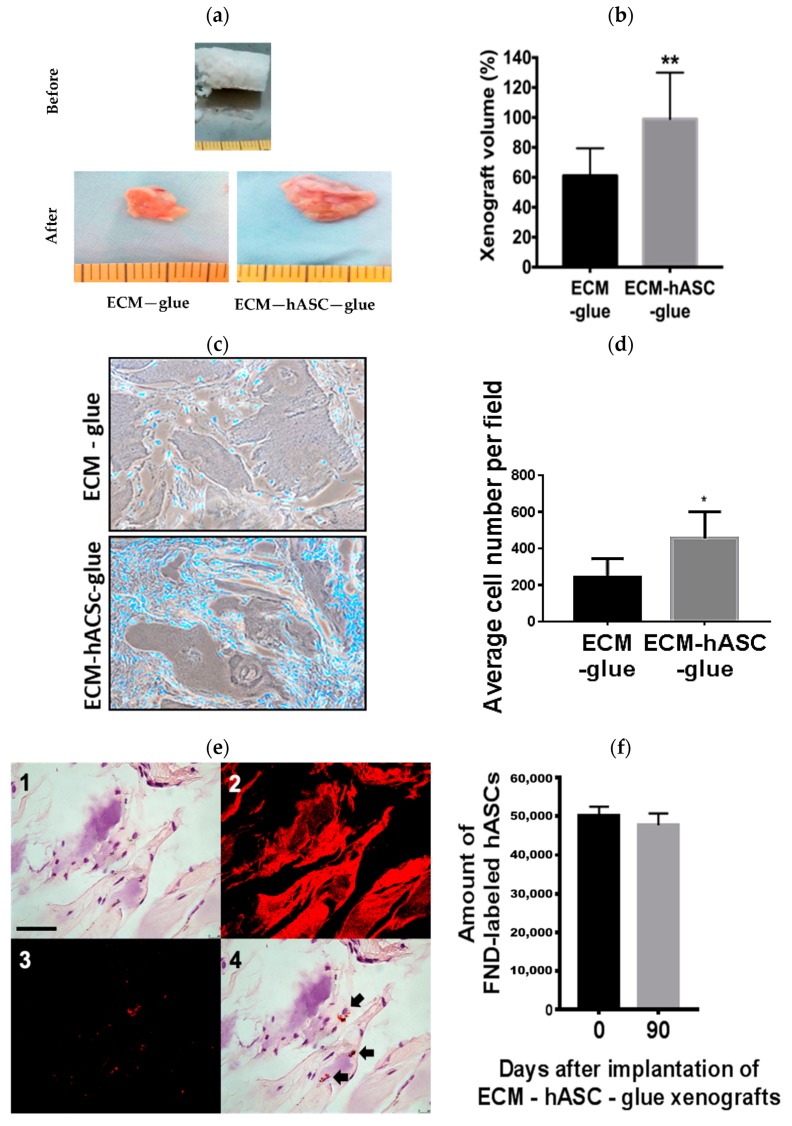
Clinically available materials, including allogeneic irradiated costal cartilage and fibrin glue polymer, were used as scaffolds for in vivo chondrogenic differentiation of human adipose-derived stem/stromal cells (hASCs) in the attempt to develop a more efficient treatment over current methods. Current studies include the use of growth-factor stimulation, tissue engineering, and biocompatible materials; however, most methods involve complicated processes and pose clinical limitations. In this report, the xenografts in the experimental group composed of a diced decellularized cartilage extracellular matrix (ECM), hASCs, and fibrin glue polymer were implanted into the subcutaneous layer of nude mice, and the results were compared with two groups of controls; one control group received implantation of decellularized cartilage ECM and fibrin glue polymer, and the other control group received implantation of hASCs mixed with fibrin glue polymer. To evaluate whether hASCs had in vivo chondrogenesis in the xenografts, hASCs were labeled with fluorescent nanodiamonds (FNDs), a biocompatible and photostable nanomaterial, to allow for long-term detection and histological analysis. Increased cellularity, glycosaminoglycan, and collagen deposition were found by the histological examination in the experimental group compared with control groups. With the background-free detection technique and time-gated fluorescence imaging, the numbers and locations of the FND-labeled hASCs could be detected by confocal microscopy. The chondrocyte-specific markers, such as aggrecan and type II collagen, were colocalized with cells containing signals of FNDs which indicated in vivo chondrogenesis of hASCs. Taken together, functional in vivo chondrogenesis of the hASCs could be achieved by clinically available decellularized cartilage ECM and fibrin glue polymer in the nude mice model without in vitro chondrogenic induction. The fluorescent signals of FNDs in hASCs can be detected in histological analysis, such as hematoxylin and eosin staining (H&E staining) without the interference of the autofluorescence. Our study may warrant future clinical applications of the combination of decellular cartilage ECM, fibrin glue polymer, and hASCs for cartilage repair.
临床可用材料,包括异体辐照肋软骨和纤维蛋白胶聚合物,被用作人脂肪来源的干/基质细胞(hASC)体内软骨形成分化的支架,试图开发一种比现有方法更有效的治疗方法。目前的研究包括使用生长因子刺激、组织工程和生物相容性材料;然而,大多数方法涉及复杂的过程并存在临床局限性。在本报告中,将由切碎的脱细胞软骨细胞外基质(ECM)、hASC和纤维蛋白胶聚合物组成的实验组异种移植物植入裸鼠皮下层,并将结果与两组对照组进行比较;一个对照组接受脱细胞软骨ECM和纤维蛋白胶聚合物的植入,另一个对照组接受hASC与纤维蛋白胶聚合物混合的植入。为了评估hASC在异种移植物中是否发生体内软骨形成,用荧光纳米金刚石(FND)标记hASC,FND是一种生物相容性和光稳定性纳米材料,用于长期检测和组织学分析。与对照组相比,实验组通过组织学检查发现细胞增多、糖胺聚糖和胶原蛋白沉积增加。利用无背景检测技术和时间门控荧光成像,通过共聚焦显微镜可以检测到FND标记的hASC的数量和位置。软骨细胞特异性标志物,如聚集蛋白聚糖和II型胶原蛋白,与含有FND信号的细胞共定位,这表明hASC发生了体内软骨形成。综上所述,在裸鼠模型中,无需体外软骨形成诱导,临床可用的脱细胞软骨ECM和纤维蛋白胶聚合物即可实现hASC的功能性体内软骨形成。在组织学分析中,如苏木精和伊红染色(H&E染色),可以检测到hASC中FND的荧光信号,而不受自发荧光的干扰。我们的研究可能为脱细胞软骨ECM、纤维蛋白胶聚合物和hASC联合用于软骨修复的未来临床应用提供依据。