Cunningham Rachel A, Holland Martha, McWilliams Emily, Hodi Frank Stephen, Severgnini Mariano
Department of Medical Oncology, Center for Immuno-Oncology, Dana-Farber Cancer Institute, 450 Brookline, Ave Mayer Building 305, Boston, MA 02215, USA.
J Biol Methods. 2019 Jun 3;6(2):e114. doi: 10.14440/jbm.2019.283. eCollection 2019.
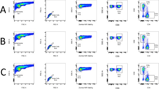
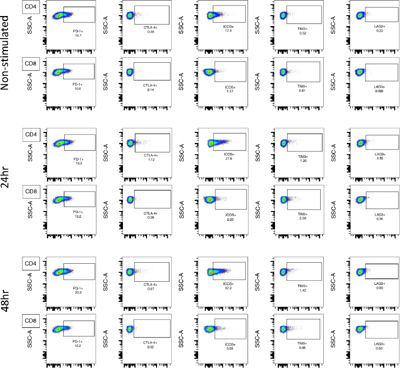
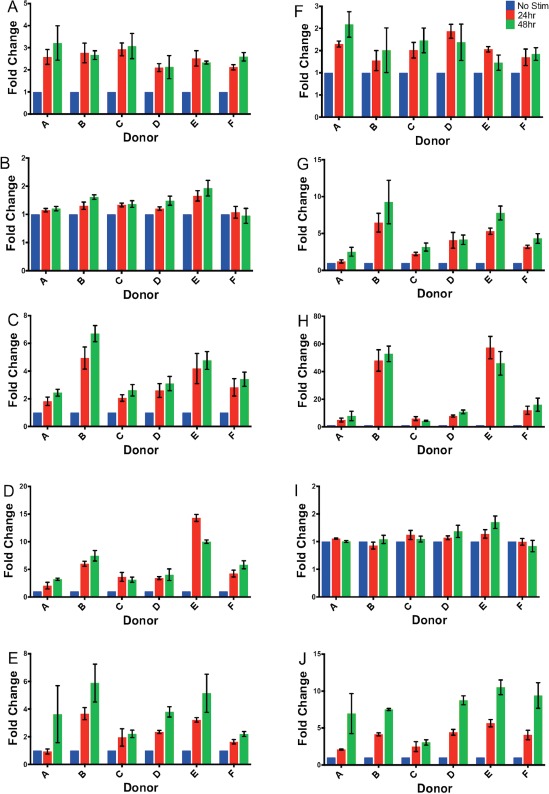
As checkpoint inhibitor immunotherapies gain traction among cancer researchers and clinicians, the need grows for assays that can definitively phenotype patient immune cells. Herein, we present an 8-color flow cytometry panel for lineage and immune checkpoint markers and validate it using healthy human donor peripheral blood mononuclear cells (PBMCs). Flow cytometry data was generated on a BD LSR Fortessa and supported by Luminex multiplex soluble immunoassay. Our data showed significant variation between donors at both baseline and different stages of activation, as well as a trend in increasing expression of checkpoint markers on stimulated CD4 and CD8 T-cells with time. Soluble immune checkpoint quantification assays revealed that LAG-3, TIM-3, CTLA-4, and PD-1 soluble isoforms are upregulated after stimulation. This 8-color flow cytometry panel, supported here by soluble immunoassay, can be used to identify and evaluate immune checkpoints on T-lymphocytes in cryopreserved human PBMC samples. This panel is ideal for characterizing checkpoint expression in clinical samples for which cryopreservation is necessary.
随着检查点抑制剂免疫疗法在癌症研究人员和临床医生中越来越受到关注,对能够明确鉴定患者免疫细胞表型的检测方法的需求也日益增加。在此,我们展示了一种用于谱系和免疫检查点标志物的8色流式细胞术检测方案,并使用健康人类供体的外周血单核细胞(PBMC)对其进行了验证。流式细胞术数据是在BD LSR Fortessa上生成的,并得到了Luminex多重可溶性免疫测定的支持。我们的数据显示,在基线和不同激活阶段,供体之间存在显著差异,并且随着时间的推移,刺激后的CD4和CD8 T细胞上检查点标志物的表达有增加的趋势。可溶性免疫检查点定量检测显示,刺激后LAG-3、TIM-3、CTLA-4和PD-1可溶性异构体上调。这种由可溶性免疫测定支持的8色流式细胞术检测方案可用于鉴定和评估冷冻保存的人类PBMC样本中T淋巴细胞上的免疫检查点。该检测方案非常适合用于对需要冷冻保存的临床样本中的检查点表达进行表征。