Oddo Letizia, Paradossi Gaio, Cerroni Barbara, Ben-Harush Carmit, Ariel Eti, Di Meco Francesco, Ram Zvi, Grossman Rachel
Dipartimento di Scienze e Tecnologie Chimiche, Universitá degli Studi di Roma Tor Vergata, 00133 Roma, Italy.
Department of Neurosurgery, Tel Aviv Medical Center, affiliated to the Sackler Faculty of Medicine, Tel-Aviv University, 6997801 Tel-Aviv, Israel.
ACS Omega. 2019 Aug 8;4(8):13371-13381. doi: 10.1021/acsomega.9b01544. eCollection 2019 Aug 20.
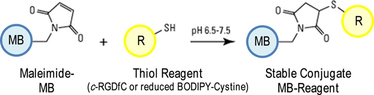
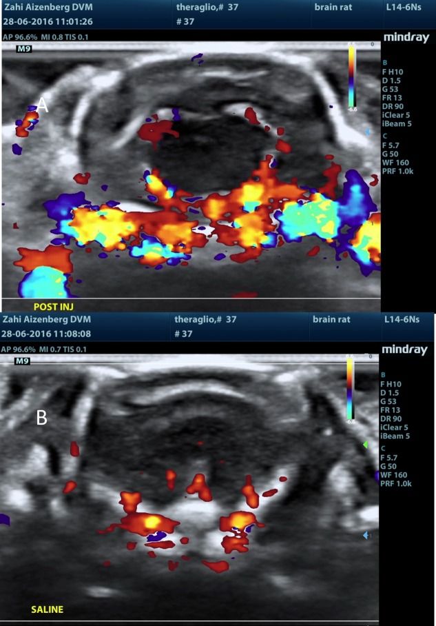
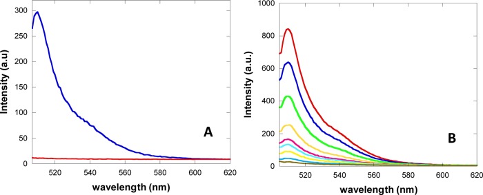
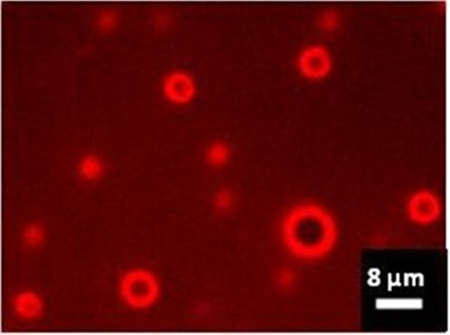
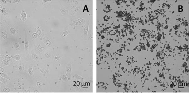
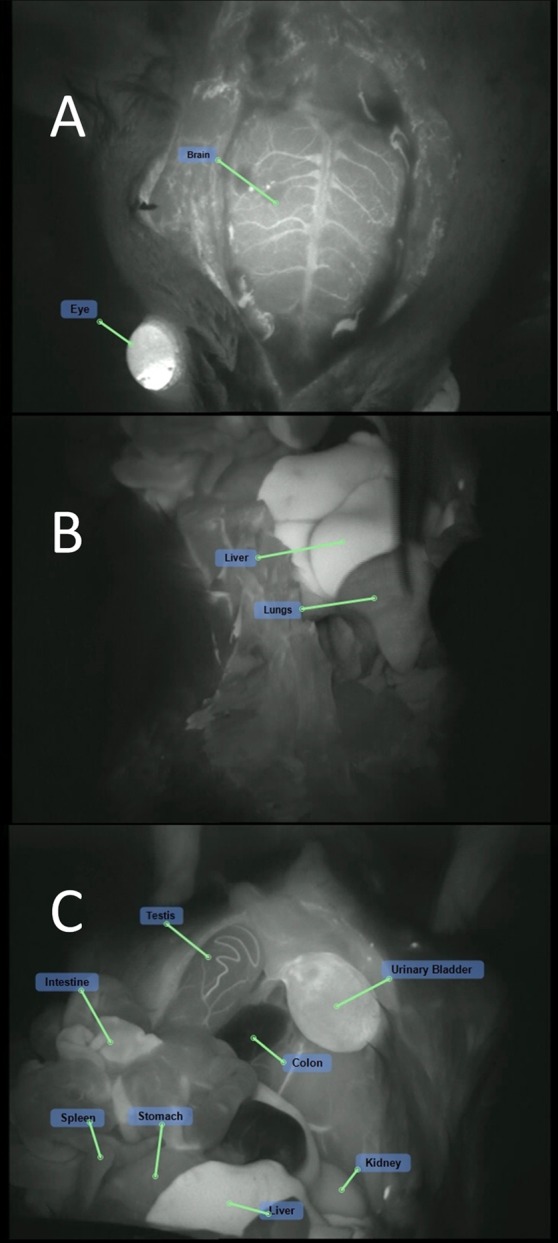
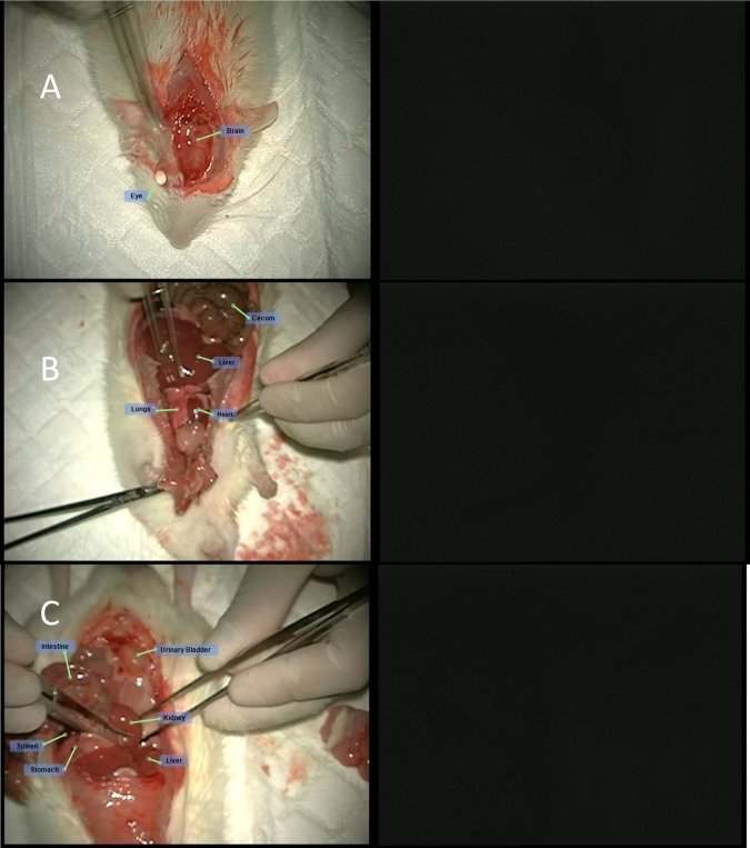
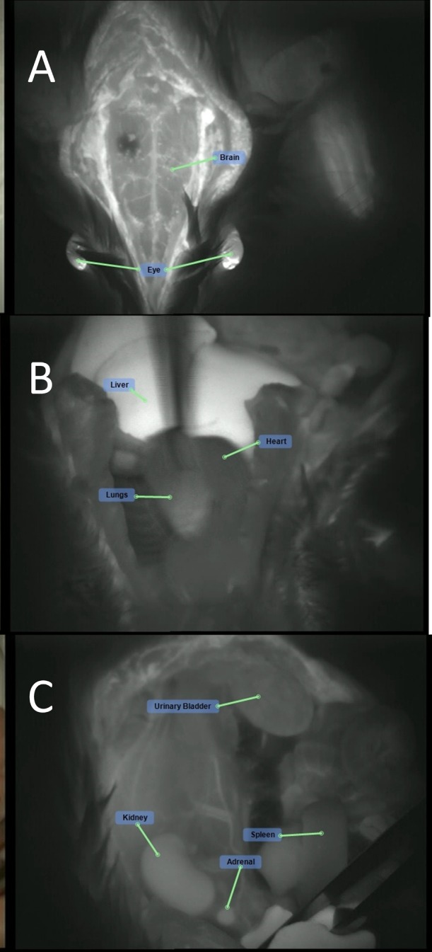
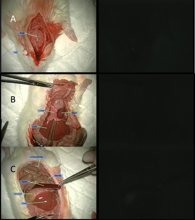
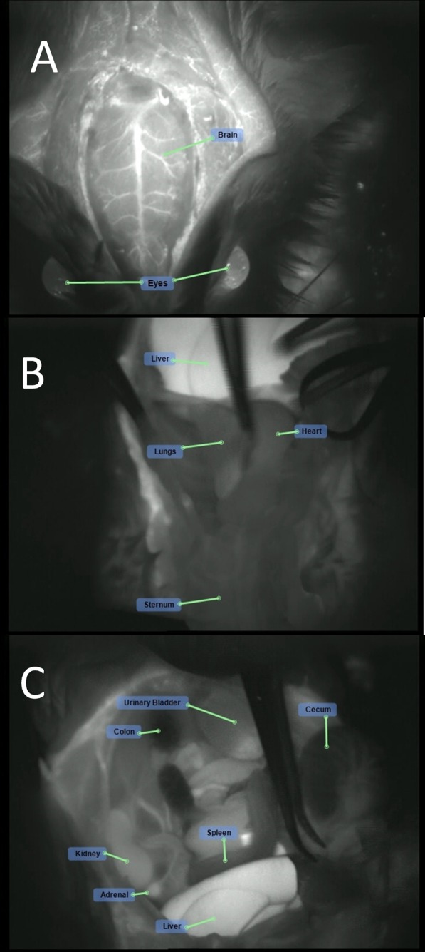
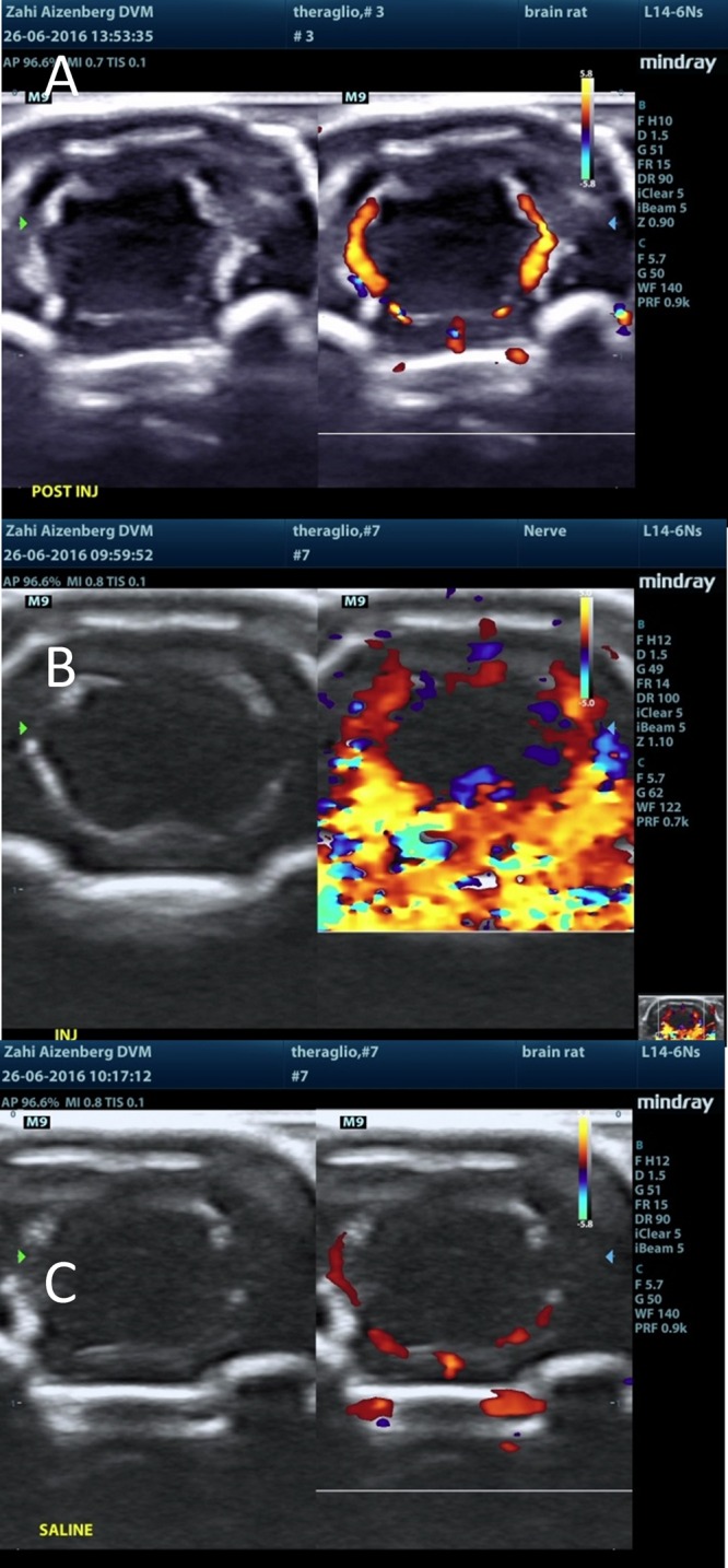
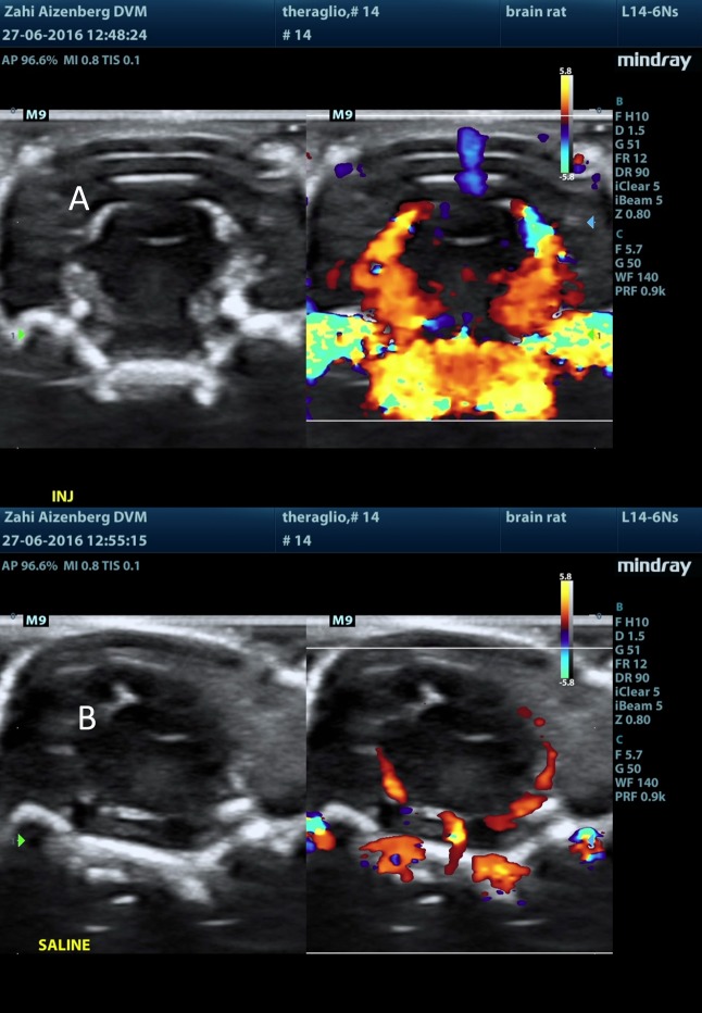
Maximal resection of intrinsic brain tumors is a major prognostic factor for survival. Real-time intraoperative imaging tools, including ultrasound (US), are crucial for maximal resection of such tumors. Microbubbles (MBs) are clinically used in daily practice as a contrast agent for ultrasound and can be further developed to serve combined therapeutic and diagnostic purposes. To achieve this goal, we have developed novel MBs conjugated to specific ligands to receptors which are overexpressed in brain tumors. These MBs are designed to target a tumor tissue, visualize it, and deliver therapeutic molecules into it. The objective of this study was to assess the biodistribution of the test items: We used MBs labeled with indocyanine green (MB-ICG) for visualization and MBs conjugated to a cyclic molecule containing the tripeptide Arg-Gly-Asp (RGD) labeled with ICG (MB-RGD-ICG) to target brain tumor integrins as the therapeutic tools. Male Sprague Dawley rats received a single dose of each MB preparation. The identification of the MB in various organs was monitored by fluorescence microscopy in anesthetized animals as well as real-time US for brain imaging. Equally sized control groups under identical conditions were used in this study. One control group was used to establish fluorescence background conditions (ICG), and two control groups were used to test autofluorescence from the test items (MBs and MB-RGD). ICG with or without MBs (naked or RGD-modified) was detected in the brain vasculature and also in other organs. The pattern, duration, and intensity of the fluorescence signal could not be differentiated between animals treated with ICG alone and animals treated with microbubbles MBs-ICG or MBs-RGD-ICG. Following MB injection, either naked or combined with RGD, there was a sharp rise in the Doppler signal within seconds of injection in the brain. The signal was mainly located at the choroid plexus, septum pellucidum, and the meninges of the brain. The signal subsided within a few minutes. Injection of saline or ICG alone to respective animals did not result in a similar raised signal. Following a single intravenous administration of MB-ICG and MB-RGD-ICG to rats, the MBs were found to be effectively present in the brain.
脑内原发性肿瘤的最大程度切除是影响生存的主要预后因素。包括超声(US)在内的实时术中成像工具对于此类肿瘤的最大程度切除至关重要。微泡(MBs)在日常临床实践中用作超声造影剂,并且可以进一步开发以用于联合治疗和诊断目的。为实现这一目标,我们开发了与在脑肿瘤中过表达的受体的特异性配体偶联的新型微泡。这些微泡旨在靶向肿瘤组织、使其可视化并将治疗分子递送至其中。本研究的目的是评估受试物的生物分布:我们使用用吲哚菁绿标记的微泡(MB-ICG)进行可视化,并使用与含三肽精氨酸-甘氨酸-天冬氨酸(RGD)的环状分子偶联并用ICG标记的微泡(MB-RGD-ICG)靶向脑肿瘤整合素作为治疗工具。雄性Sprague Dawley大鼠接受每种微泡制剂的单剂量注射。通过荧光显微镜在麻醉动物中监测微泡在各个器官中的识别情况,并通过实时超声进行脑成像。本研究使用了在相同条件下大小相同的对照组。一个对照组用于建立荧光背景条件(ICG),两个对照组用于测试受试物(微泡和MB-RGD)的自发荧光。在脑血管系统以及其他器官中检测到了含有或不含有微泡(裸微泡或RGD修饰微泡)的ICG。单独用ICG处理的动物与用微泡MBs-ICG或MBs-RGD-ICG处理的动物之间,荧光信号的模式、持续时间和强度无法区分。注射微泡后,无论是否与RGD结合,脑内注射后数秒内多普勒信号都会急剧上升。信号主要位于脉络丛、透明隔和脑膜。信号在几分钟内消退。向相应动物注射生理盐水或单独的ICG不会导致类似的信号升高。对大鼠单次静脉注射MB-ICG和MB-RGD-ICG后,发现微泡有效地存在于脑中。