Department of Oral and Maxillofacial Surgery/Central Laboratory, Peking University School and Hospital of Stomatology, Beijing, 100081, China.
Laboratory of Biomaterials and Regenerative Medicine, Academy for Advanced Interdisciplinary Studies, Peking University, Beijing, 100871, China.
Stem Cell Res Ther. 2019 Aug 29;10(1):276. doi: 10.1186/s13287-019-1378-7.
Tooth loss caused by caries or injuries has a negative effect on human health; thus, it is important to develop a reliable method of tooth regeneration. Research on tooth regeneration has mainly focused on mouse pluripotent stem cells, mouse embryonic stem cells, and adult stem cells from various sources in mice, whereas little has examined the differentiation of human embryonic stem (hES) cells into dental epithelium (DE) and odontogenic potential in vivo.
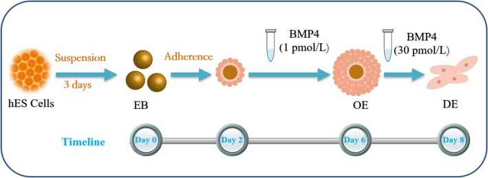
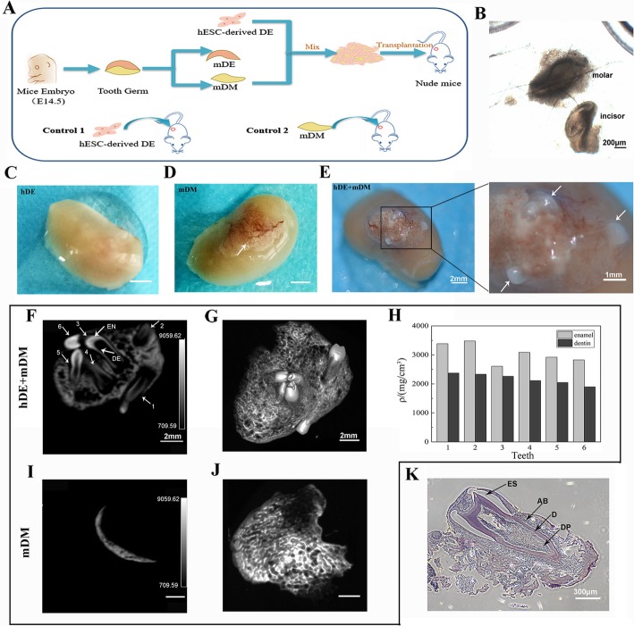
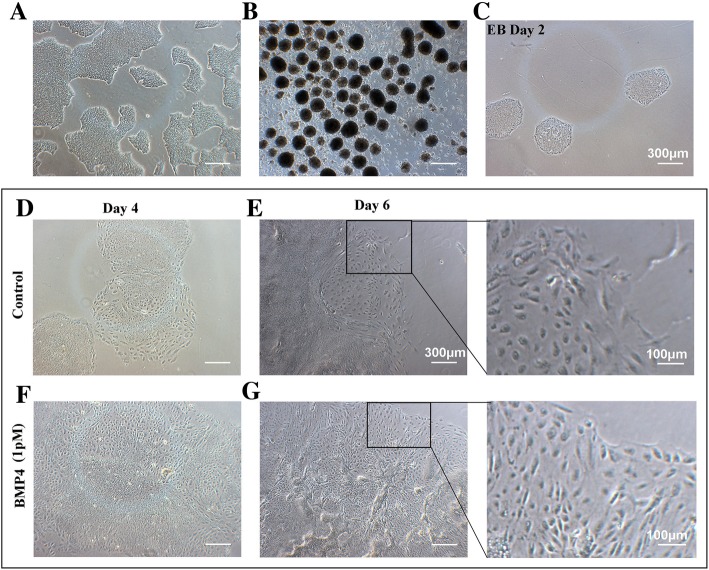
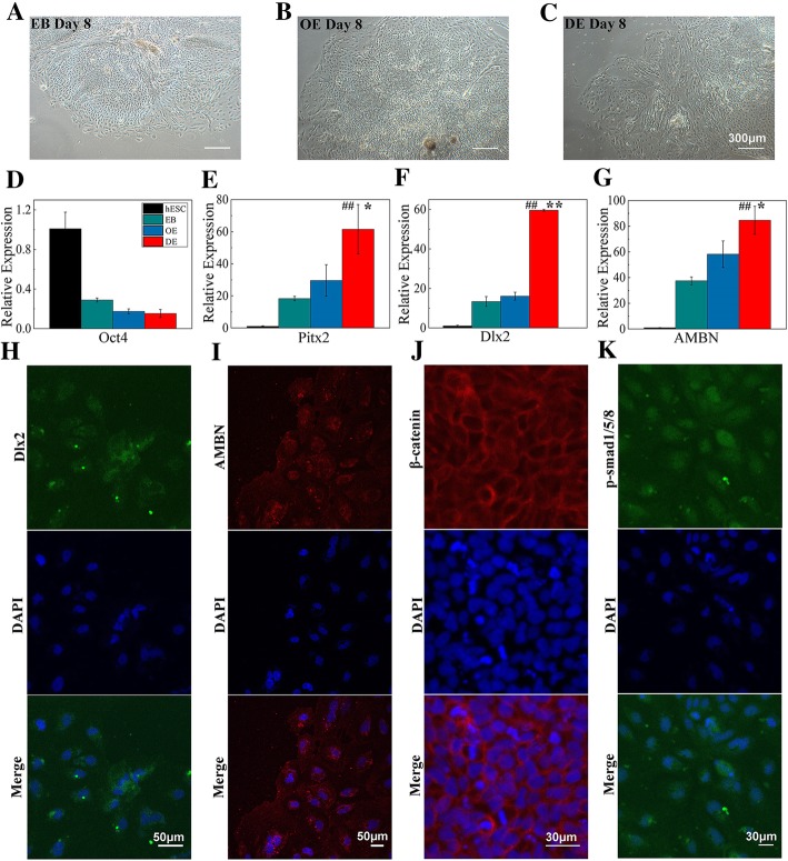
In this study, we induced hES cells to differentiate into dental epithelium using different concentrations of bone morphogenetic protein 4 (BMP4). With 1 pM BMP4, the hES cells differentiated into oral ectoderm (OE). These cells were then stimulated with 30 pM BMP4. Quantitative reverse transcription-polymerase chain reaction (qRT-PCR) and immunofluorescence showed the differentiation of OE and DE. The DE generated was mixed with embryonic day 14.5 mouse dental mesenchyme (DM) and transplanted into the renal capsules of nude mice. Thirty days later, the resulting tooth-like structures were examined by micro-computed tomography and hematoxylin and eosin staining.
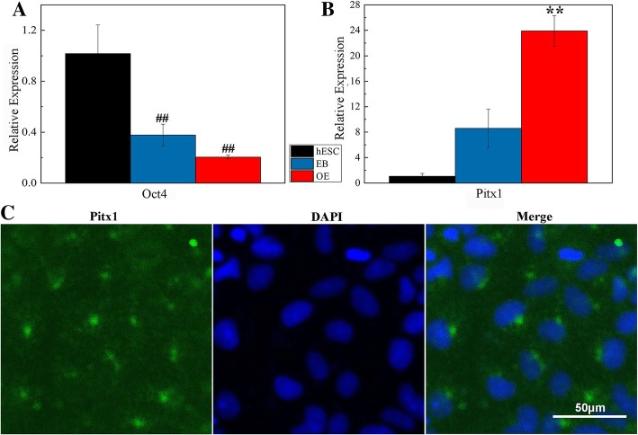
After 4 days of 1 pM BMP4 stimulation, Pitx1-positive OE formed. qRT-PCR and immunofluorescence revealed that induction with 30 pM BMP4 for 2 days caused the OE to differentiate into Pitx2/Dlx2/AMBN-positive DE-like cells. These cells also expressed β-catenin and p-Smad1/5/8, which are key proteins in the Wnt/β-catenin and Bmp signaling pathways, respectively. Thirty days after in vivo transplantation, six teeth with enamel and dentin had formed on the kidney.
These results show that hES cells differentiated into DE after sequential stimulation with different concentrations of BMP4, and the DE thus generated showed odontogenic potential.
龋病或外伤导致的牙齿缺失对人类健康有负面影响,因此,开发一种可靠的牙齿再生方法非常重要。牙齿再生的研究主要集中在小鼠多能干细胞、小鼠胚胎干细胞和各种来源的成年干细胞上,而很少研究人类胚胎干细胞(hES)细胞在体内向牙上皮(DE)分化和牙原性潜能。
在这项研究中,我们使用不同浓度的骨形态发生蛋白 4(BMP4)诱导 hES 细胞分化为牙上皮。用 1 pM BMP4 诱导 hES 细胞分化为口腔外胚层(OE)。然后用 30 pM BMP4 刺激这些细胞。定量逆转录聚合酶链反应(qRT-PCR)和免疫荧光显示 OE 和 DE 的分化。生成的 DE 与胚胎第 14.5 天的小鼠牙间质(DM)混合,并移植到裸鼠的肾囊中。30 天后,通过微计算机断层扫描和苏木精和伊红染色检查所得的牙样结构。
在 1 pM BMP4 刺激 4 天后,形成了 Pitx1 阳性的 OE。qRT-PCR 和免疫荧光显示,用 30 pM BMP4 诱导 2 天可使 OE 分化为 Pitx2/Dlx2/AMBN 阳性的 DE 样细胞。这些细胞还表达β-catenin 和 p-Smad1/5/8,它们分别是 Wnt/β-catenin 和 Bmp 信号通路中的关键蛋白。体内移植 30 天后,在肾脏上形成了 6 颗具有釉质和牙本质的牙齿。
这些结果表明,hES 细胞在受到不同浓度 BMP4 的顺序刺激后分化为 DE,由此产生的 DE 显示出牙原性潜能。