• 文献检索
  • 文档翻译
  • 深度研究
  • 学术资讯
  • Suppr Zotero 插件Zotero 插件
  • 邀请有礼
  • 套餐&价格
  • 历史记录
应用&插件
Suppr Zotero 插件Zotero 插件浏览器插件Mac 客户端Windows 客户端微信小程序
定价
高级版会员购买积分包购买API积分包
服务
文献检索文档翻译深度研究API 文档MCP 服务
关于我们
关于 Suppr公司介绍联系我们用户协议隐私条款
关注我们

Suppr 超能文献

核心技术专利:CN118964589B侵权必究
粤ICP备2023148730 号-1Suppr @ 2026

文献检索

告别复杂PubMed语法,用中文像聊天一样搜索,搜遍4000万医学文献。AI智能推荐,让科研检索更轻松。

立即免费搜索

文件翻译

保留排版,准确专业,支持PDF/Word/PPT等文件格式,支持 12+语言互译。

免费翻译文档

深度研究

AI帮你快速写综述,25分钟生成高质量综述,智能提取关键信息,辅助科研写作。

立即免费体验

色素性视网膜炎一种变异型中的双侧视网膜血管瘤样增殖

Bilateral Retinal Angiomatous Proliferation in a Variant of Retinitis Pigmentosa.

作者信息

Aloe G, De Sanctis C M, Strafella C, Cascella R, Missiroli F, Cesareo M, Giardina E, Ricci F

机构信息

Unit Retinal Pathology PTV Foundation, Tor Vergata University, Rome, Italy.

Department of Biomedicine and Prevention, Tor Vergata University, Rome, Italy.

出版信息

Case Rep Ophthalmol Med. 2019 Jul 31;2019:8547962. doi: 10.1155/2019/8547962. eCollection 2019.

DOI:10.1155/2019/8547962
PMID:31467752
原文链接:https://pmc.ncbi.nlm.nih.gov/articles/PMC6699319/
Abstract

PURPOSE

To describe the first case of bilateral retinal angiomatous proliferation (RAP) in a patient with a variant of retinitis pigmentosa (RP).

CASE REPORT

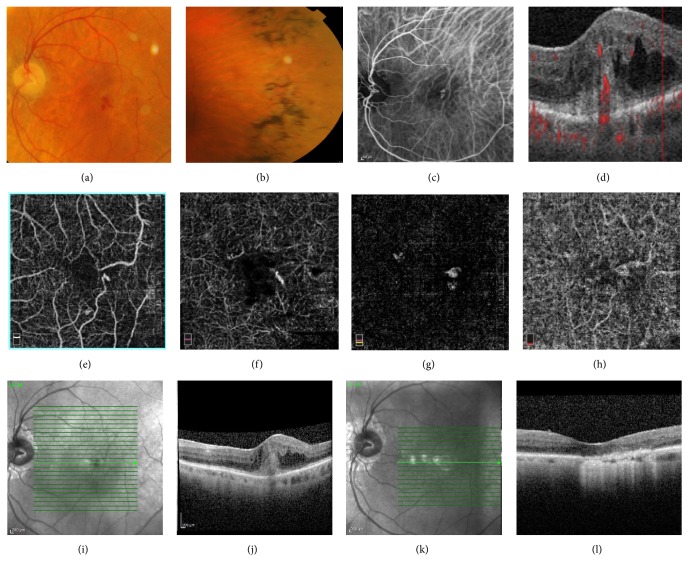
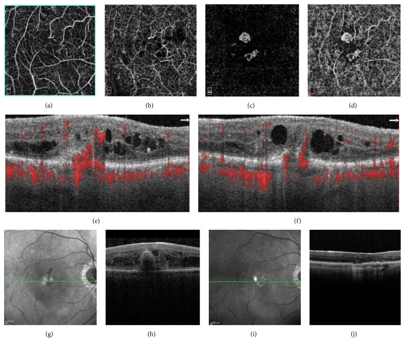
An 85-year-old man with RP presented with visual acuity decrease and metamorphopsia in the left eye (LE). Fundus examination revealed typical signs of RP in both eyes, associated with intraretinal macular hemorrhage in the LE. Multimodal imaging, using Colour fundus Photography, Fluorescein (FA), and Indocyanine Green Angiography (ICGA) as well as Spectral-Domain Optical Coherence Tomography (SD-OCT) and Optical Coherence Tomography Angiography (OCTA), revealed a type 3 neovascular lesion in the involved eye. Genetic testing (NGS analysis) was performed to search for genetic variants correlated with the disease phenotype displayed by the patient. The patient was treated with intravitreal injections of bevacizumab, according to a fixed protocol of bimonthly injections plus a booster dose at second month. After 9 months, he was referred for visual acuity decrease and metamorphopsia in the fellow eye, where SD-OCT/OCTA showed a type 3 neovascular lesion in the right eye (RE). He was scheduled for intravitreal injections of bevacizumab. In both eyes, treatment with intravitreal bevacizumab was successful.

摘要

目的

描述一名患有色素性视网膜炎(RP)变异型患者的首例双侧视网膜血管瘤样增生(RAP)病例。

病例报告

一名85岁的RP男性患者,左眼(LE)出现视力下降和视物变形。眼底检查发现双眼均有典型的RP体征,左眼伴有视网膜黄斑部出血。采用彩色眼底照相、荧光素(FA)、吲哚菁绿血管造影(ICGA)以及光谱域光学相干断层扫描(SD-OCT)和光学相干断层扫描血管造影(OCTA)等多模态成像技术,发现患眼存在3型新生血管病变。进行了基因检测(NGS分析)以寻找与患者所表现疾病表型相关的基因变异。根据每两个月注射一次并在第二个月追加一剂的固定方案,对该患者进行了玻璃体腔注射贝伐单抗治疗。9个月后,他因对侧眼出现视力下降和视物变形前来就诊,SD-OCT/OCTA显示右眼(RE)有3型新生血管病变。他被安排进行玻璃体腔注射贝伐单抗治疗。双眼玻璃体腔注射贝伐单抗治疗均取得成功。

https://cdn.ncbi.nlm.nih.gov/pmc/blobs/5f9a/6699319/3d5f2569b25d/CRIOPM2019-8547962.002.jpg
https://cdn.ncbi.nlm.nih.gov/pmc/blobs/5f9a/6699319/7822aa3cc334/CRIOPM2019-8547962.001.jpg
https://cdn.ncbi.nlm.nih.gov/pmc/blobs/5f9a/6699319/3d5f2569b25d/CRIOPM2019-8547962.002.jpg
https://cdn.ncbi.nlm.nih.gov/pmc/blobs/5f9a/6699319/7822aa3cc334/CRIOPM2019-8547962.001.jpg
https://cdn.ncbi.nlm.nih.gov/pmc/blobs/5f9a/6699319/3d5f2569b25d/CRIOPM2019-8547962.002.jpg

相似文献

1
Bilateral Retinal Angiomatous Proliferation in a Variant of Retinitis Pigmentosa.色素性视网膜炎一种变异型中的双侧视网膜血管瘤样增殖
Case Rep Ophthalmol Med. 2019 Jul 31;2019:8547962. doi: 10.1155/2019/8547962. eCollection 2019.
2
Retinal Angiomatous Proliferation in a Patient with Retinitis Pigmentosa.视网膜色素变性患者的视网膜血管瘤增生。
Genes (Basel). 2023 Jul 13;14(7):1438. doi: 10.3390/genes14071438.
3
IMPROVED DETECTION AND DIAGNOSIS OF POLYPOIDAL CHOROIDAL VASCULOPATHY USING A COMBINATION OF OPTICAL COHERENCE TOMOGRAPHY AND OPTICAL COHERENCE TOMOGRAPHY ANGIOGRAPHY.采用光学相干断层扫描和光学相干断层扫描血管造影相结合的方法提高息肉状脉络膜血管病变的检测和诊断。
Retina. 2019 Sep;39(9):1655-1663. doi: 10.1097/IAE.0000000000002228.
4
OPTICAL COHERENCE TOMOGRAPHY ANGIOGRAPHY IMAGING OF CHOROIDAL NEOVASCULARIZATION SECONDARY TO CHOROIDAL RUPTURE TREATED BY INTRAVITREAL RANIBIZUMAB.经玻璃体腔内雷珠单抗治疗的脉络膜破裂所致脉络膜新生血管的光相干断层扫描血管造影成像。
Retin Cases Brief Rep. 2022 Mar 1;16(2):222-225. doi: 10.1097/ICB.0000000000000932.
5
Retinal Angiomatous Proliferation: Multimodal Imaging Characteristics and Follow-up with Eye-Tracked Spectral Domain Optical Coherence Tomography of Precursor Lesions.视网膜血管瘤样增生:多模态成像特征及对前驱病变的眼跟踪光谱域光学相干断层扫描随访
Turk J Ophthalmol. 2018 Apr;48(2):66-69. doi: 10.4274/tjo.03780. Epub 2018 Apr 25.
6
SWEPT-SOURCE OPTICAL COHERENCE TOMOGRAPHY ANGIOGRAPHY FINDINGS IN A CASE OF PACHYCHOROID NEOVASCULOPATHY IN RETINITIS PIGMENTOSA.特发性脉络膜新生血管性色素性视网膜炎中厚脉络膜新生血管的扫频源光学相干断层扫描血管造影研究。
Retin Cases Brief Rep. 2022 May 1;16(3):300-304. doi: 10.1097/ICB.0000000000000962. Epub 2020 Jan 6.
7
Type 3 Neovascularization Associated with Retinitis Pigmentosa.与色素性视网膜炎相关的3型新生血管形成
Case Rep Ophthalmol. 2017 Apr 10;8(1):245-249. doi: 10.1159/000471790. eCollection 2017 Jan-Apr.
8
OCT-angiography assessing quiescent and active choroidal neovascularization in retinitis pigmentosa associated with pathogenic variant.光学相干断层扫描血管造影评估与致病变异相关的色素性视网膜炎中的静止和活动性脉络膜新生血管形成。
Eur J Ophthalmol. 2022 Jul;32(4):NP98-NP102. doi: 10.1177/11206721211004396. Epub 2021 Mar 18.
9
Outcomes in Eyes with Retinal Angiomatous Proliferation in the Comparison of Age-Related Macular Degeneration Treatments Trials (CATT).年龄相关性黄斑变性治疗试验(CATT)中视网膜血管瘤样增生患者的治疗结果
Ophthalmology. 2016 Mar;123(3):609-16. doi: 10.1016/j.ophtha.2015.10.034. Epub 2015 Dec 8.
10
Optic Coherence Tomography Angiography Findings of Bilateral Choroidal Neovascularization Associated with Optic Disc Drusen Treated with Intravitreal Aflibercept Injection.玻璃体腔注射阿柏西普治疗与视盘玻璃疣相关的双侧脉络膜新生血管的光学相干断层扫描血管造影结果
Case Rep Ophthalmol Med. 2020 Mar 10;2020:8398054. doi: 10.1155/2020/8398054. eCollection 2020.

引用本文的文献

1
The potential key role of choroidal non-perfusion and rod degeneration in the pathogenesis of macular neovascularization type 3.脉络膜无灌注和杆状细胞变性在 3 型黄斑新生血管发病机制中的潜在关键作用。
Eye (Lond). 2024 Jul;38(10):1836-1839. doi: 10.1038/s41433-024-03034-z. Epub 2024 Mar 18.
2
Retinal Angiomatous Proliferation in a Patient with Retinitis Pigmentosa.视网膜色素变性患者的视网膜血管瘤增生。
Genes (Basel). 2023 Jul 13;14(7):1438. doi: 10.3390/genes14071438.
3
The RAP study, report 4: morphological and topographical characteristics of multifocal macular neovascularization type 3.

本文引用的文献

1
Non-syndromic retinitis pigmentosa.非综合征性视网膜色素变性。
Prog Retin Eye Res. 2018 Sep;66:157-186. doi: 10.1016/j.preteyeres.2018.03.005. Epub 2018 Mar 27.
2
Uncovering genetic and non-genetic biomarkers specific for exudative age-related macular degeneration: significant association of twelve variants.揭示渗出性年龄相关性黄斑变性特有的遗传和非遗传生物标志物:十二个变异体的显著关联
Oncotarget. 2017 Dec 12;9(8):7812-7821. doi: 10.18632/oncotarget.23241. eCollection 2018 Jan 30.
3
Towards the application of precision medicine in Age-Related Macular Degeneration.
RAP 研究,报告 4:多灶性黄斑新生血管病变 3 型的形态学和解剖学特征。
Graefes Arch Clin Exp Ophthalmol. 2022 Jan;260(1):141-147. doi: 10.1007/s00417-021-05332-8. Epub 2021 Aug 26.
迈向精准医学在年龄相关性黄斑变性中的应用。
Prog Retin Eye Res. 2018 Mar;63:132-146. doi: 10.1016/j.preteyeres.2017.11.004. Epub 2017 Nov 29.
4
Assessing individual risk for AMD with genetic counseling, family history, and genetic testing.使用遗传咨询、家族史和基因检测评估 AMD 的个体风险。
Eye (Lond). 2018 Feb;32(2):446-450. doi: 10.1038/eye.2017.192. Epub 2017 Sep 15.
5
Type 3 Neovascularization Associated with Retinitis Pigmentosa.与色素性视网膜炎相关的3型新生血管形成
Case Rep Ophthalmol. 2017 Apr 10;8(1):245-249. doi: 10.1159/000471790. eCollection 2017 Jan-Apr.
6
Optical coherence tomography (OCT) in retinal angiomatous proliferation (RAP).视网膜血管瘤样增生(RAP)中的光学相干断层扫描(OCT)。
Eur J Ophthalmol. 2002;12(6):467-472. doi: 10.5301/EJO.2008.2283.
7
The role of Sema4A in angiogenesis, immune responses, carcinogenesis, and retinal systems.Sema4A在血管生成、免疫反应、致癌作用及视网膜系统中的作用。
Cell Adh Migr. 2016 Nov;10(6):692-699. doi: 10.1080/19336918.2016.1215785. Epub 2016 Oct 13.
8
OPTICAL COHERENCE TOMOGRAPHY ANGIOGRAPHY IN EARLY TYPE 3 NEOVASCULARIZATION.光学相干断层扫描血管造影术在早期3型新生血管形成中的应用
Retina. 2015 Nov;35(11):2236-41. doi: 10.1097/IAE.0000000000000834.
9
The Genetics and the Genomics of Primary Congenital Glaucoma.原发性先天性青光眼的遗传学与基因组学
Biomed Res Int. 2015;2015:321291. doi: 10.1155/2015/321291. Epub 2015 Sep 16.
10
Comparative analysis between saliva and buccal swabs as source of DNA: lesson from HLA-B*57:01 testing.唾液与口腔拭子作为DNA来源的比较分析:来自HLA-B*57:01检测的经验教训。
Pharmacogenomics. 2015;16(10):1039-46. doi: 10.2217/pgs.15.59. Epub 2015 Jul 31.