Sayadi Jihene, Miere Alexandra, Souied Eric H, Cohen Salomon Y
aDepartment of Ophthalmology, Intercity Hospital and University Paris Est, Créteil, France.
bOphthalmic Center for Imaging and Laser, Paris, France.
Case Rep Ophthalmol. 2017 Apr 10;8(1):245-249. doi: 10.1159/000471790. eCollection 2017 Jan-Apr.
To report a case of type 3 neovascular lesion in a patient with retinitis pigmentosa (RP) complicated by macular edema.
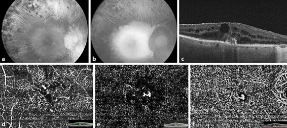
A 78-year-old man with a long follow-up for RP was referred for painless visual acuity decrease in the right eye. Best-corrected visual acuity was 20/125 in the right eye and 20/40 in the left. Fundus examination showed typical RP and macular edema in both eyes. In the right eye, spectral domain optical coherence tomography revealed a marked cystic macular edema associated with disruption of the Bruch membrane/retinal pigment epithelium complex overlying a pigmentary epithelium detachment, with a vascular structure which appeared to originate from the deep capillary plexus and to be connected with the subretinal pigment epithelium space. Optical coherence tomography angiography showed a high-flow vessel infiltrating the outer retinal layers in the deep capillary plexus segmentation, and a tuft-shaped, bright, high-flow network that seemed to be connected with the subretinal pigment epithelium space in the outer retinal layer segmentation. This presentation was consistent with an early type 3 neovascular lesion in the right eye.
Type 3 neovascularization may be considered a possible complication of RP.
报告一例视网膜色素变性(RP)合并黄斑水肿患者发生3型新生血管病变的病例。
一名长期随访的78岁男性因右眼无痛性视力下降前来就诊。右眼最佳矫正视力为20/125,左眼为20/40。眼底检查显示双眼典型的RP和黄斑水肿。在右眼,光谱域光学相干断层扫描显示黄斑区有明显的囊样水肿,伴有覆盖色素上皮脱离处的布鲁赫膜/视网膜色素上皮复合体的破坏,有一个血管结构似乎起源于深层毛细血管丛并与视网膜下色素上皮间隙相连。光学相干断层扫描血管造影显示在深层毛细血管丛节段有一条高流量血管浸润外层视网膜,在外层视网膜节段有一个簇状、明亮、高流量的网络,似乎与视网膜下色素上皮间隙相连。此表现符合右眼早期3型新生血管病变。
3型新生血管化可能被视为RP的一种潜在并发症。