Department of Orthopedic Surgery, Nara Medical University, Shijocho 840, Kashihara, Nara, 634-8522, Japan.
Department of Artificial Joint and Regenerative Medicine for Bone and Cartilage, Nara Medical University, Shijocho 840, Kashihara, Nara, 634-8522, Japan.
BMC Musculoskelet Disord. 2019 Aug 31;20(1):396. doi: 10.1186/s12891-019-2777-8.
Treatment of anterior cruciate ligament injuries commonly involves the use of polyethylene terephthalate (PET) artificial ligaments for reconstruction. However, the currently available methods require long fixation periods, thereby necessitating the development of alternative methods to accelerate the healing process between tendons and bones. Thus, we developed and evaluated a novel technique that utilizes silicate-substituted strontium (SrSiP).
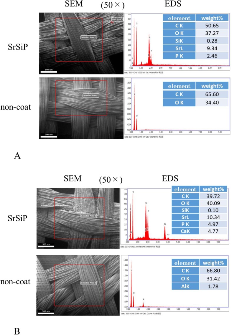
PET films, nano-coated with SrSiP, were prepared. Bone marrow mesenchymal cells (BMSCs) from femurs of male rats were cultured and seeded at a density of 1.0 × 10/cm onto the SrSiP-coated and non-coated PET film, and subsequently placed in an osteogenic medium. The osteocalcin concentration secreted into the medium was compared in each case. Next, PET artificial ligament, nano-coated with SrSiP, were prepared. BMSCs were seeded at a density of 4.5 × 10/cm onto the SrSiP-coated, and non-coated artificial ligament, and then placed in osteogenic medium. The osteocalcin and calcium concentrations in the culture medium were measured on the 8th, 10th, 12th, and 14th day of culture. Furthermore, mRNA expression of osteocalcin, alkaline phosphatase (ALP), bone morphogenetic protein-2 (BMP2), and runt-related transcription factor 2 (Runx2) was evaluated by qPCR. We transplanted the SrSiP-coated and non-coated artificial ligament to the tibiae of mature New Zealand white rabbits. Two months later, we sacrificed them and histologically evaluated them.
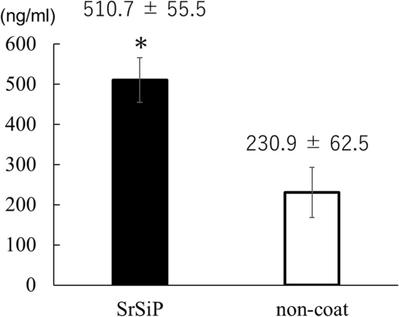
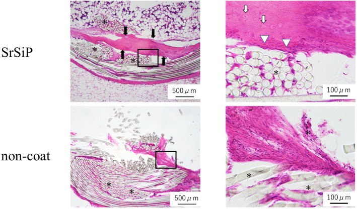
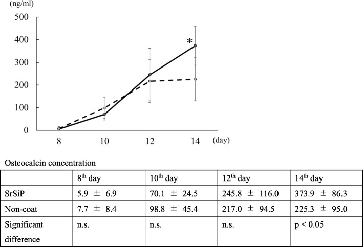
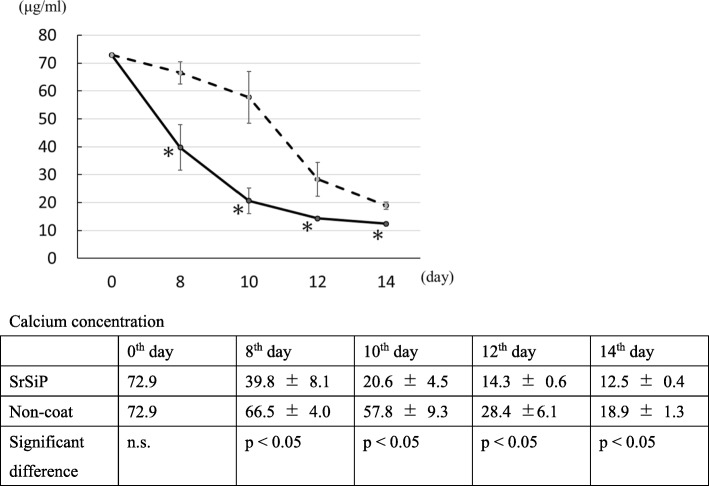
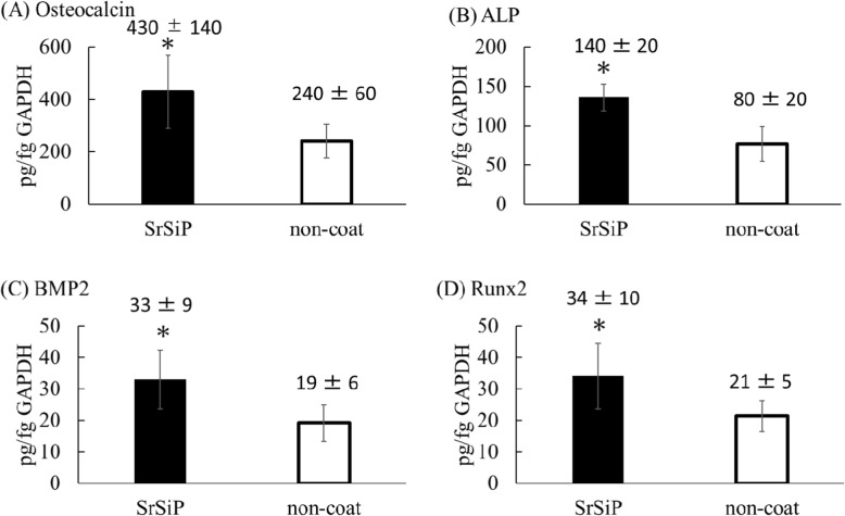
The secretory osteocalcin concentration in the medium on the film was significantly higher for the SrSiP group than for the non-coated group. Secretory osteocalcin concentration in the medium on the artificial ligament was also significantly higher in the SrSiP group than in the non-coated group on the 14th day. Calcium concentration on the artificial ligament was significantly lower in the SrSiP group than in the non-coated group on the 8th, 10th, 12th, and 14th day. In qPCR as well, OC, ALP, BMP2, and Runx2 mRNA expression were significantly higher in the SrSiP group than in the non-coated group. Newly formed bone was histologically found around the artificial ligament in the SrSiP group.
Our findings demonstrate that artificial ligaments using SrSiP display high osteogenic potential and thus may be efficiently used in future clinical applications.
前交叉韧带损伤的治疗通常涉及使用聚对苯二甲酸乙二醇酯(PET)人工韧带进行重建。然而,目前可用的方法需要长时间的固定期,因此需要开发替代方法来加速肌腱和骨骼之间的愈合过程。因此,我们开发并评估了一种利用硅取代锶(SrSiP)的新方法。
制备了纳米涂层 SrSiP 的 PET 薄膜。从雄性大鼠股骨中培养骨髓间充质细胞(BMSCs),以 1.0×10/cm 的密度接种到 SrSiP 涂层和非涂层的 PET 薄膜上,然后将其置于成骨培养基中。比较每种情况下分泌到培养基中的骨钙素浓度。接下来,制备纳米涂层 SrSiP 的 PET 人工韧带。将 BMSCs 以 4.5×10/cm 的密度接种到 SrSiP 涂层和非涂层的人工韧带,然后将其置于成骨培养基中。在培养的第 8、10、12 和 14 天测量培养基中骨钙素和钙的浓度。此外,通过 qPCR 评估骨钙素、碱性磷酸酶(ALP)、骨形态发生蛋白-2(BMP2)和 runt 相关转录因子 2(Runx2)的 mRNA 表达。我们将 SrSiP 涂层和非涂层的人工韧带移植到成熟新西兰白兔的胫骨中。两个月后,将它们处死并进行组织学评估。
薄膜上 SrSiP 组的培养基中分泌的骨钙素浓度明显高于非涂层组。在第 14 天,SrSiP 组的人工韧带培养基中分泌的骨钙素浓度也明显高于非涂层组。在第 8、10、12 和 14 天,人工韧带中 SrSiP 组的钙浓度明显低于非涂层组。在 qPCR 中,SrSiP 组的 OC、ALP、BMP2 和 Runx2 mRNA 表达也明显高于非涂层组。在 SrSiP 组中,在人工韧带周围发现了新形成的骨。
我们的研究结果表明,使用 SrSiP 的人工韧带具有较高的成骨潜力,因此可能在未来的临床应用中得到有效利用。