Kawasaki Sachiko, Inagaki Yusuke, Akahane Manabu, Furukawa Akira, Shigematsu Hideki, Tanaka Yasuhito
Department of Orthopaedic Surgery, Nara Medical University, Shijocho 840, Kashihara, Nara, 634-8522, Japan.
Department of Health and Welfare Services, National Institute of Public Health, South 2-3-6, Wako, Saitama, 351-0197, Japan.
BMC Musculoskelet Disord. 2020 Oct 19;21(1):692. doi: 10.1186/s12891-020-03716-1.
Polyether-ether-ketone (PEEK) is increasingly being used for spinal applications. However, because of its biologically inactive nature, there are risks of false joint loosening and sinking. PEEK materials are coated with apatite to enhance the osteoconductive properties. In this study, we aimed to evaluate whether strontium apatite stimulate osteogenesis on the surface of PEEK by using the CO laser technique.
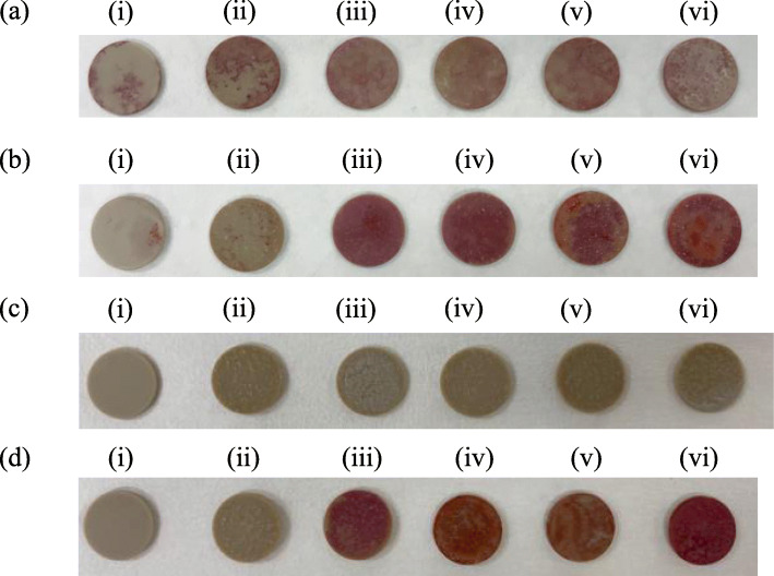
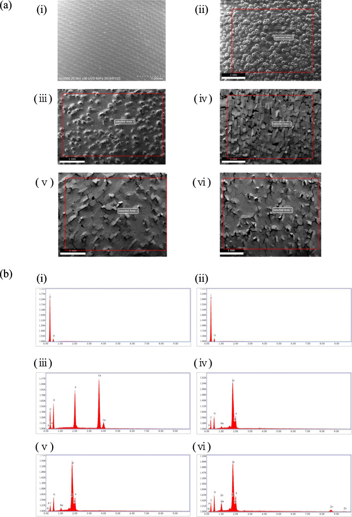
We prepared non-coated disks, laser-exposed disks without apatite, and four types of apatite-coated by laser PEEK disks (hydroxyapatite (HAP), strontium hydroxyapatite (SrHAP), silicate-substituted strontium apatite (SrSiP), and silicate-zinc-substituted strontium apatite (SrZnSiP)). A part of the study objective was testing various types of apatite coatings. Bone marrow mesenchymal cells (BMSCs) of rats were seeded at a density of 2 × 10/cm onto each apatite-coated, non-coated, and laser-irradiated PEEK disks. The disks were then placed in osteogenic medium, and alkaline phosphatase (ALP) staining and Alizarin red staining of BMSCs grown on PEEK disks were performed after 14 days of culture. The concentrations of osteocalcin (OC) and calcium in the culture medium were measured on days 8 and 14 of cell culture. Furthermore, mRNA expression of osteocalcin, ALP, runt-related transcription factor 2 (Runx2), collagen type 1a1 (Col1a1), and collagen type 4a1 (Col4a1) was evaluated by qPCR.
The staining for ALP and Alizarin red S was more strongly positive on the apatite-coated PEEK disks compared to that on non-coated or laser-exposed without coating PEEK disks. The concentration of osteocalcin secreted into the medium was also significantly higher in case of the SrHAP, SrSiP, and SrZnSiP disks than that in the case of the non-coated on day14. The calcium concentration in the PEEK disk was significantly lower in all apatite-coated disks than that in the pure PEEK disks on day 14. In qPCR, OC and ALP mRNA expression was significantly higher in the SrZnSiP disks than that in the pure PEEK disks.
Our findings demonstrate that laser bonding of apatite-along with trace elements-on the PEEK disk surfaces might provide the material with surface property that enable better osteogenesis.
聚醚醚酮(PEEK)越来越多地用于脊柱应用。然而,由于其生物惰性,存在假关节松动和下沉的风险。PEEK材料涂覆有磷灰石以增强骨传导性能。在本研究中,我们旨在通过使用CO激光技术评估锶磷灰石是否能刺激PEEK表面的骨生成。
我们制备了未涂覆的圆盘、未涂覆磷灰石的激光照射圆盘以及四种通过激光涂覆磷灰石的PEEK圆盘(羟基磷灰石(HAP)、锶羟基磷灰石(SrHAP)、硅酸盐取代的锶磷灰石(SrSiP)和硅酸盐 - 锌取代的锶磷灰石(SrZnSiP))。该研究的部分目标是测试各种类型的磷灰石涂层。将大鼠骨髓间充质细胞(BMSCs)以2×10⁶/cm²的密度接种到每个涂覆有磷灰石、未涂覆和激光照射的PEEK圆盘上。然后将圆盘置于成骨培养基中,培养14天后对在PEEK圆盘上生长的BMSCs进行碱性磷酸酶(ALP)染色和茜素红染色。在细胞培养的第8天和第14天测量培养基中骨钙素(OC)和钙的浓度。此外,通过qPCR评估骨钙素、ALP、 runt相关转录因子2(Runx2)、1a1型胶原蛋白(Col1a1)和4a1型胶原蛋白(Col4a1)的mRNA表达。
与未涂覆或未涂覆磷灰石的激光照射PEEK圆盘相比,涂覆有磷灰石的PEEK圆盘上的ALP和茜素红S染色阳性更强。在第14天,SrHAP、SrSiP和SrZnSiP圆盘分泌到培养基中的骨钙素浓度也显著高于未涂覆的圆盘。在第14天,所有涂覆有磷灰石的圆盘上PEEK圆盘中的钙浓度均显著低于纯PEEK圆盘。在qPCR中,SrZnSiP圆盘中OC和ALP mRNA表达显著高于纯PEEK圆盘。
我们的研究结果表明,在PEEK圆盘表面进行磷灰石与微量元素的激光结合可能为材料提供能够实现更好骨生成的表面特性。