Department of Pathology, Tianjin First Central Hospital, Number 24, Convalescent Road, Nankai District, Tianjin 300192, China.
Dis Markers. 2019 Aug 5;2019:9436047. doi: 10.1155/2019/9436047. eCollection 2019.
MCOLN1 (mucolipin subfamily, member 1) was first identified as an autophagic regulator, which was essential for efficient fusion of both autophagosomes and late endosomes with lysosomes. This study is aimed at investigating the role of MCOLN1 in the development of pancreatic ductal adenocarcinoma (PDAC).
Immunohistochemistry (IHC) assay was conducted to evaluate the expression level of MCOLN1 in 82 human PDAC tumor tissues. Overall survival (OS) and recurrence-free survival (RFS) analysis was performed to assess the prognosis of patients. Colony formation and MTT assays [3-(4,5-dimethyl-2-thiazolyl)-2,5-diphenyl-2-H-tetrazolium bromide] were performed to measure the proliferation capacity of tumor cells. The expression level of related genes was measured by RT-PCR (reverse transcription polymerase chain reaction) and western blot assays. The animal model was used to examine the effects of indicated protein on tumorigenesis in vivo.
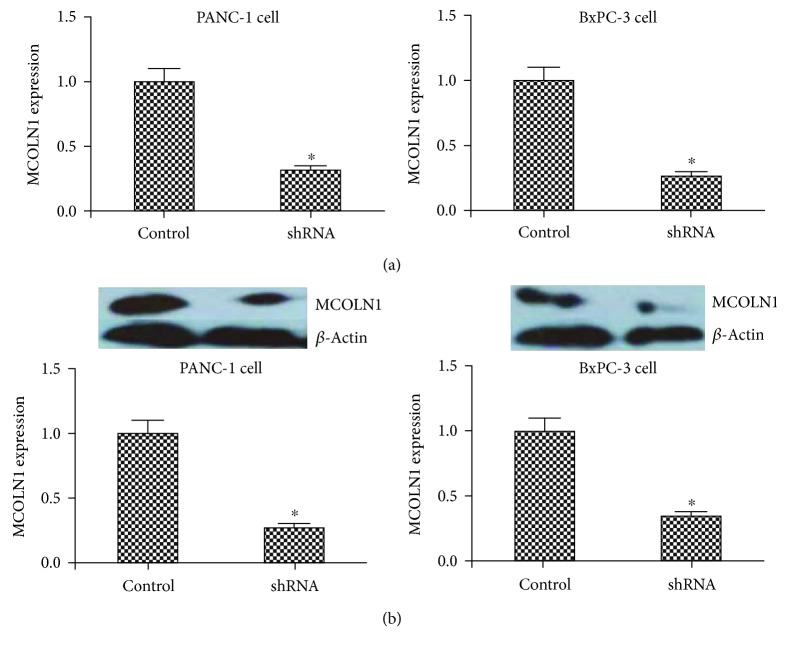
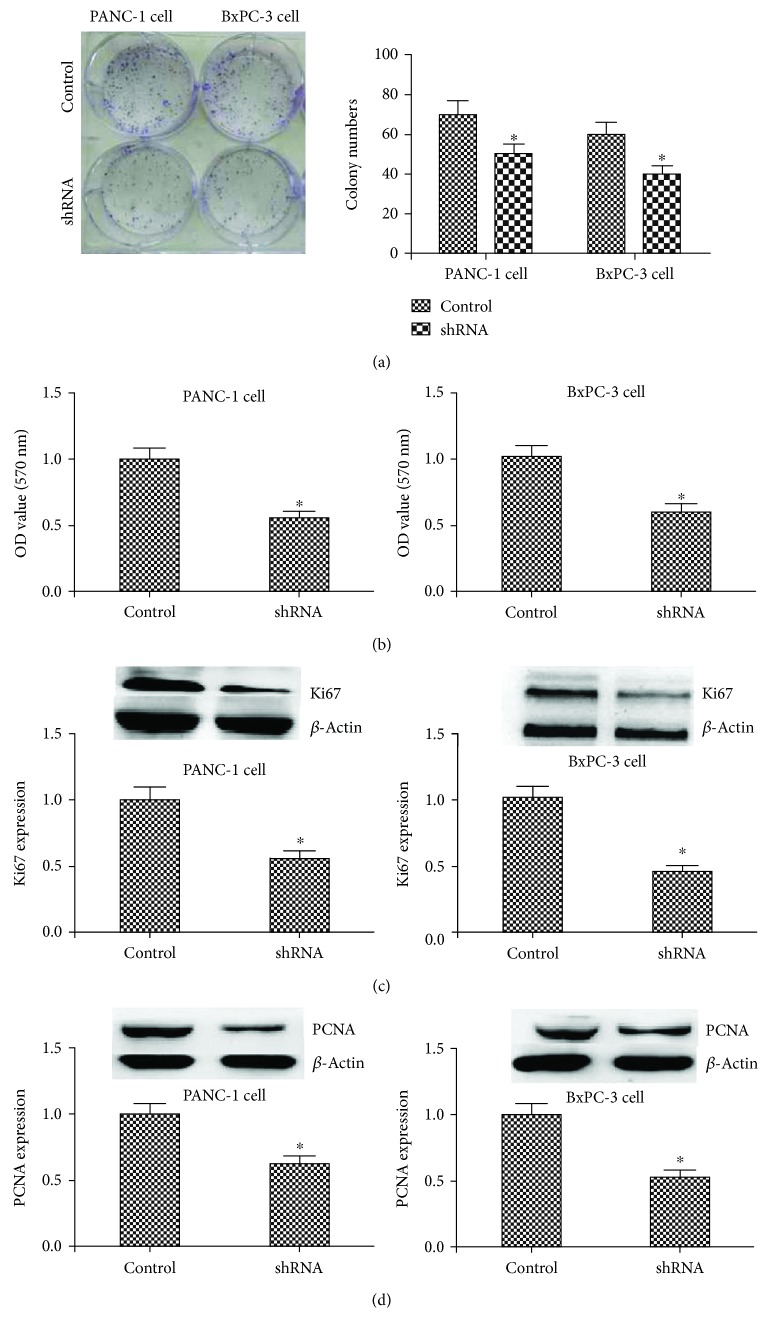
The results of IHC showed that a high level of MCOLN1 expression was associated with the poor clinical characteristics of PDAC patients. OS and RFS were significantly worse in patients with high MCOLN1 expression. Silencing of MCOLN1 dramatically blocked the proliferation of PDAC cells. Mechanism studies confirmed that knockdown of MCOLN1 decreased the expression of Ki67 and PCNA (proliferating cell nuclear antigen), two markers of cell proliferation. In vivo, MCOILN1 depletion reduced the formation and growth of tumors in mice.
The high level of MCOLN1 expression was associated with poor clinical outcomes of PDAC patients. MCOLN1 ablation could inhibit PDAC proliferation of both in vitro and in vivo, which provide a new insight and novel therapeutic target for the treatment of PDAC.
MCOLN1(多噬菌素亚家族成员 1)最初被鉴定为自噬调节剂,对于自噬体和晚期内体与溶酶体的有效融合至关重要。本研究旨在探讨 MCOLN1 在胰腺导管腺癌(PDAC)发展中的作用。
采用免疫组织化学(IHC)检测 82 例人 PDAC 肿瘤组织中 MCOLN1 的表达水平。进行总生存(OS)和无复发生存(RFS)分析以评估患者的预后。通过集落形成和 MTT 测定[3-(4,5-二甲基-2-噻唑基)-2,5-二苯基-2-H-四唑溴盐]来测量肿瘤细胞的增殖能力。通过 RT-PCR(逆转录聚合酶链反应)和 Western blot 测定来测量相关基因的表达水平。利用动物模型来检测指示蛋白对体内肿瘤发生的影响。
IHC 结果表明,MCOLN1 高表达与 PDAC 患者的不良临床特征相关。高 MCOLN1 表达的患者 OS 和 RFS 明显更差。沉默 MCOLN1 可显著阻断 PDAC 细胞的增殖。机制研究证实,下调 MCOLN1 可降低 Ki67 和 PCNA(增殖细胞核抗原)的表达,这是细胞增殖的两个标志物。体内研究中,MCOILN1 耗竭减少了小鼠肿瘤的形成和生长。
MCOLN1 高表达与 PDAC 患者的不良临床结局相关。MCOLN1 缺失可抑制 PDAC 的体外和体内增殖,为 PDAC 的治疗提供了新的见解和新的治疗靶点。