Department of Radiology & Nuclear Medicine, Maastricht University Medical Centre, Maastricht, the Netherlands.
Department of Neurology, Maastricht University Medical Centre, Maastricht, the Netherlands.
J Magn Reson Imaging. 2020 Apr;51(4):1170-1180. doi: 10.1002/jmri.26920. Epub 2019 Sep 4.
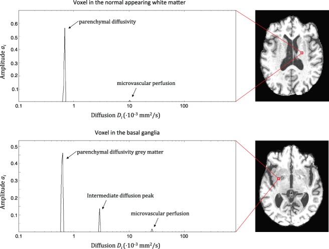
Cerebral intravoxel incoherent motion (IVIM) imaging assumes two components. However, more compartments are likely present in pathologic tissue. We hypothesized that spectral analysis using a nonnegative least-squares (NNLS) approach can detect an additional, intermediate diffusion component, distinct from the parenchymal and microvascular components, in lesion-prone regions.
To investigate the presence of this intermediate diffusion component and its relation with cerebral small vessel disease (cSVD)-related lesions.
Prospective cross-sectional study.
Patients with cSVD (n = 69, median age 69.8) and controls (n = 39, median age 68.9).
FIELD STRENGTH/SEQUENCE: Whole-brain inversion recovery IVIM acquisition at 3.0T.
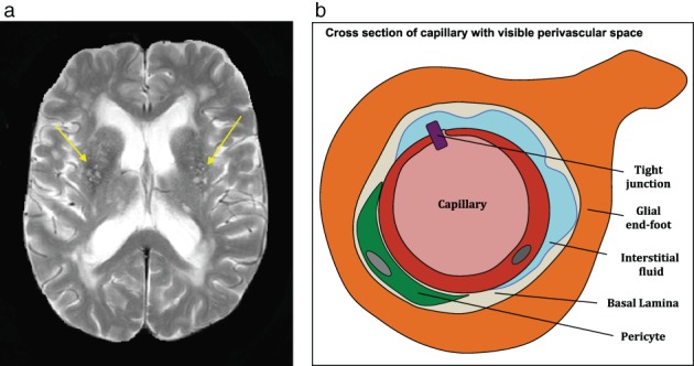
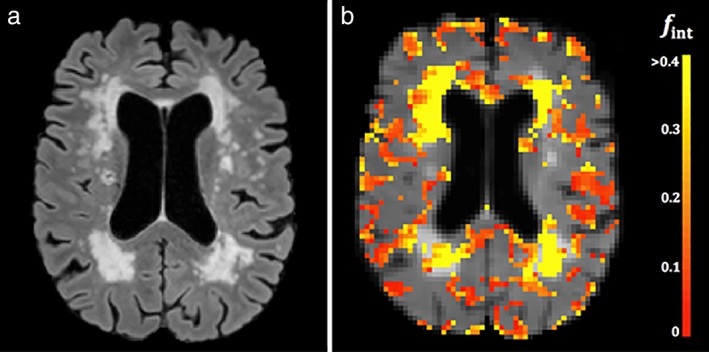
Enlarged perivascular spaces (PVS) were rated by three raters. White matter hyperintensities (WMH) were identified on a fluid attenuated inversion recovery (FLAIR) image using a semiautomated algorithm.
Relations between IVIM measures and cSVD-related lesions were studied using the Spearman's rank order correlation.
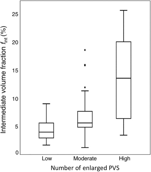
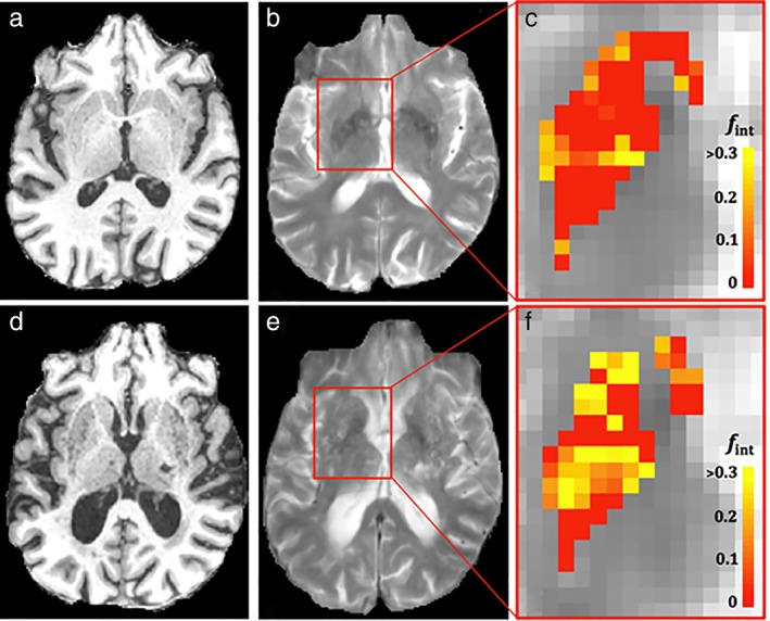
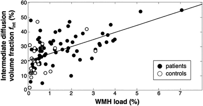
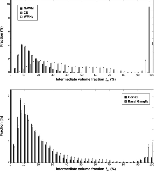
NNLS yielded diffusion spectra from which the intermediate volume fraction f was apparent between parenchymal diffusion and microvasular pseudodiffusion. WMH volume and the extent of MRI-visible enlarged PVS in the basal ganglia (BG) and centrum semiovale (CSO) were correlated with f in the WMHs, BG, and CSO, respectively. f was 4.2 ± 1.7%, 7.0 ± 4.1% and 13.6 ± 7.7% in BG and 3.9 ± 1.3%, 4.4 ± 1.4% and 4.5 ± 1.2% in CSO for the groups with low, moderate, and high number of enlarged PVS, respectively, and increased with the extent of enlarged PVS (BG: r = 0.49, P < 0.01; CSO: r = 0.23, P = 0.02). f in the WMHs was 27.1 ± 13.1%, and increased with the WMH volume (r = 0.57, P < 0.01).
We revealed the presence of an intermediate diffusion component in lesion-prone regions of cSVD and demonstrated its relation with enlarged PVS and WMHs. In tissue with these lesions, tissue degeneration or perivascular edema can lead to more freely diffusing interstitial fluid contributing to f .
2 Technical Efficacy: Stage 2 J. Magn. Reson. Imaging 2020;51:1170-1180.
脑内体素不相干运动(IVIM)成像假设存在两个分量。然而,在病变组织中可能存在更多的隔室。我们假设,使用非负最小二乘(NNLS)方法进行谱分析可以在易发病灶区域检测到与实质和微血管成分不同的额外中间扩散成分。
研究这种中间扩散成分的存在及其与脑小血管疾病(cSVD)相关病变的关系。
前瞻性横断面研究。
cSVD 患者(n=69,中位年龄 69.8)和对照组(n=39,中位年龄 68.9)。
磁场强度/序列:3.0T 全脑反转恢复 IVIM 采集。
由三位评估者评估扩大的血管周围间隙(PVS)。使用半自动算法在液体衰减反转恢复(FLAIR)图像上识别脑白质高信号(WMH)。
使用 Spearman 秩相关分析研究 IVIM 测量值与 cSVD 相关病变之间的关系。
NNLS 从扩散谱中得出中间体积分数 f,该分数在实质扩散和微血管假性扩散之间。WMH 体积和基底节(BG)和半卵圆中心(CSO)中可见的 MRI 扩大 PVS 的程度与 WMHs、BG 和 CSO 中的 f 相关。在 PVS 数量低、中、高的组中,f 在 BG 分别为 4.2±1.7%、7.0±4.1%和 13.6±7.7%,在 CSO 分别为 3.9±1.3%、4.4±1.4%和 4.5±1.2%,并随 PVS 程度的增加而增加(BG:r=0.49,P<0.01;CSO:r=0.23,P=0.02)。WMHs 中的 f 为 27.1±13.1%,并随 WMH 体积的增加而增加(r=0.57,P<0.01)。
我们在 cSVD 的易发病灶区域发现了中间扩散成分的存在,并证明了其与扩大的 PVS 和 WMHs 的关系。在存在这些病变的组织中,组织退化或血管周围水肿可能导致更多自由扩散的细胞外间质液,从而导致 f 增加。
2 技术功效:第 2 阶段 J. Magn. Reson. Imaging 2020;51:1170-1180。