Department of Medical Oncology, Amsterdam UMC, Vrije Universiteit Amsterdam/Cancer Center Amsterdam, Amsterdam, The Netherlands.
Division of Molecular Pathology, The Netherlands Cancer Institute, Amsterdam, The Netherlands.
Int J Cancer. 2020 Apr 15;146(8):2348-2359. doi: 10.1002/ijc.32668. Epub 2019 Oct 6.
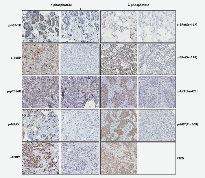
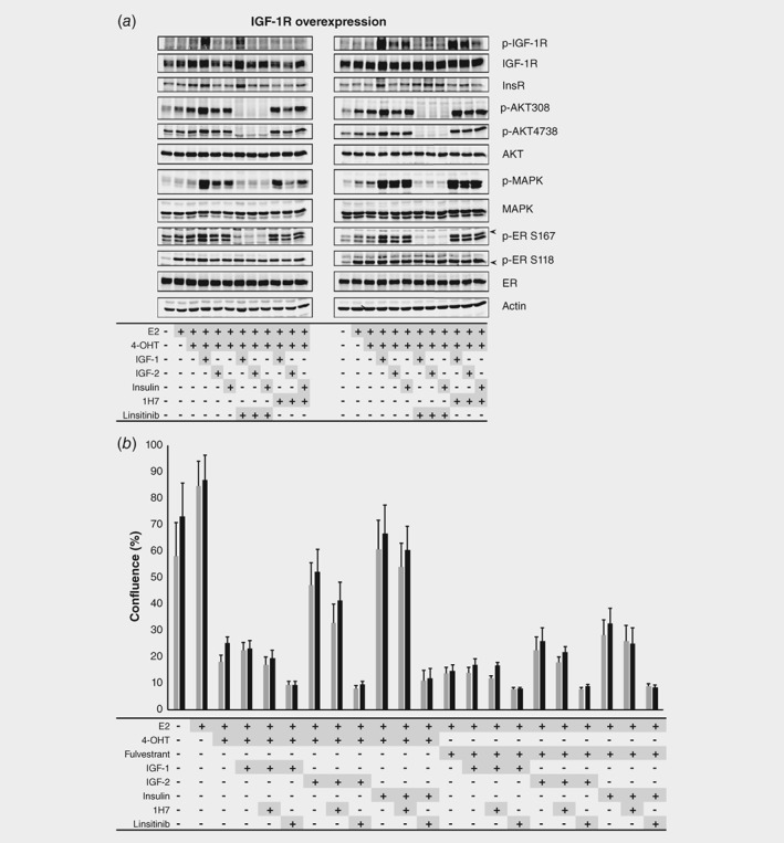
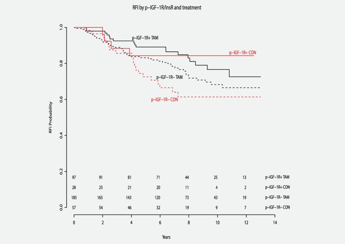
Preclinical studies indicate that activated IGF-1R can drive endocrine resistance in ER-positive (ER+) breast cancer, but its clinical relevance is unknown. We studied the effect of IGF-1R signaling on tamoxifen benefit in patients and we searched for approaches to overcome IGF-1R-mediated tamoxifen failure in cell lines. Primary tumor blocks from postmenopausal ER+ breast cancer patients randomized between adjuvant tamoxifen versus nil were recollected. Immunohistochemistry for IGF-1R, p-IGF-1R/InsR, p-ERα(Ser118), p-ERα(Ser167) and PI3K/MAPK pathway proteins was performed. Multivariate Cox models were employed to assess tamoxifen efficacy. The association between p-IGF-1R/InsR and PI3K/MAPK pathway activation in MCF-7 and T47D cells was analyzed with Western blots. Cell proliferation experiments were performed under various growth-stimulating and -inhibiting conditions. Patients with ER+, IGF-1R-positive breast cancer without p-IGF-1R/InsR staining (n = 242) had tamoxifen benefit (HR 0.41, p = 0.0038), while the results for p-IGF-1R/InsR-positive patients (n = 125) were not significant (HR 0.95, p = 0.3). High p-ERα(Ser118) or p-ERα(Ser167) expression was associated with less tamoxifen benefit. In MCF-7 cells, IGF-1R stimulation increased phosphorylation of PI3K/MAPK proteins and ERα(Ser167) regardless of IGF-1R overexpression. This could be abrogated by the dual IGF-1R/InsR inhibitor linsitinib, but not by the IGF-IR-selective antibody 1H7. In MCF-7 and T47D cells, stimulation of the IGF-1R/InsR pathway resulted in cell proliferation regardless of tamoxifen. Abrogation of cell growth was regained by addition of linsitinib. In conclusion, p-IGF-1R/InsR positivity in ER+ breast cancer is associated with reduced benefit from adjuvant tamoxifen in postmenopausal patients. In cell lines, stimulation rather than overexpression of IGF-1R is driving tamoxifen resistance to be abrogated by linsitinib.
临床前研究表明,激活的 IGF-1R 可导致 ER 阳性(ER+)乳腺癌内分泌耐药,但尚不清楚其临床相关性。我们研究了 IGF-1R 信号对 ER+乳腺癌患者他莫昔芬获益的影响,并寻找克服细胞系中 IGF-1R 介导的他莫昔芬耐药的方法。从接受辅助他莫昔芬与空白治疗的绝经后 ER+乳腺癌患者中重新采集原发肿瘤块。进行 IGF-1R、p-IGF-1R/InsR、p-ERα(Ser118)、p-ERα(Ser167)和 PI3K/MAPK 通路蛋白的免疫组织化学染色。采用多变量 Cox 模型评估他莫昔芬的疗效。用 Western blot 分析 MCF-7 和 T47D 细胞中 p-IGF-1R/InsR 与 PI3K/MAPK 通路激活之间的关联。在各种生长刺激和抑制条件下进行细胞增殖实验。无 p-IGF-1R/InsR 染色的 ER+、IGF-1R 阳性乳腺癌患者(n=242)有他莫昔芬获益(HR 0.41,p=0.0038),而 p-IGF-1R/InsR 阳性患者(n=125)的结果无统计学意义(HR 0.95,p=0.3)。高 p-ERα(Ser118)或 p-ERα(Ser167)表达与较少的他莫昔芬获益相关。在 MCF-7 细胞中,IGF-1R 刺激增加了 PI3K/MAPK 蛋白和 ERα(Ser167)的磷酸化,而不管 IGF-1R 是否过表达。这种作用可以被双重 IGF-1R/InsR 抑制剂 linsitinib 阻断,但不能被 IGF-IR 选择性抗体 1H7 阻断。在 MCF-7 和 T47D 细胞中,IGF-1R/InsR 通路的刺激导致细胞增殖,而与他莫昔芬无关。加入 linsitinib 可恢复细胞生长的抑制。总之,绝经后 ER+乳腺癌中 p-IGF-1R/InsR 阳性与辅助他莫昔芬获益减少相关。在细胞系中,IGF-1R 的刺激而非过表达导致他莫昔芬耐药,可被 linsitinib 阻断。