Department of Histopathology, NDORMS, University of Oxford, Nuffield Orthopaedic Centre, Oxford, OX3 7HE, UK.
Chemistry Research Laboratory, Mansfield Road, Oxford, OX1 3TA, UK.
J Mater Sci Mater Med. 2019 Sep 6;30(9):103. doi: 10.1007/s10856-019-6304-0.
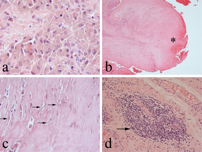
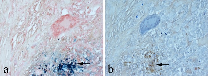
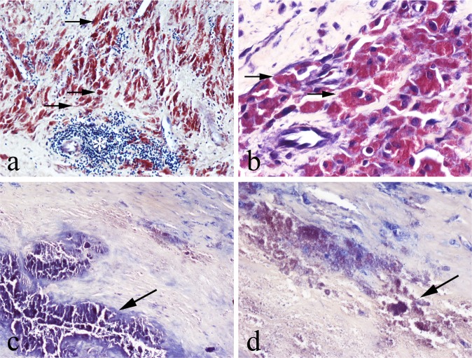
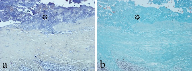
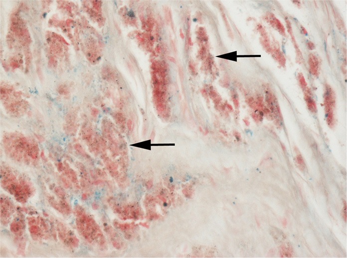
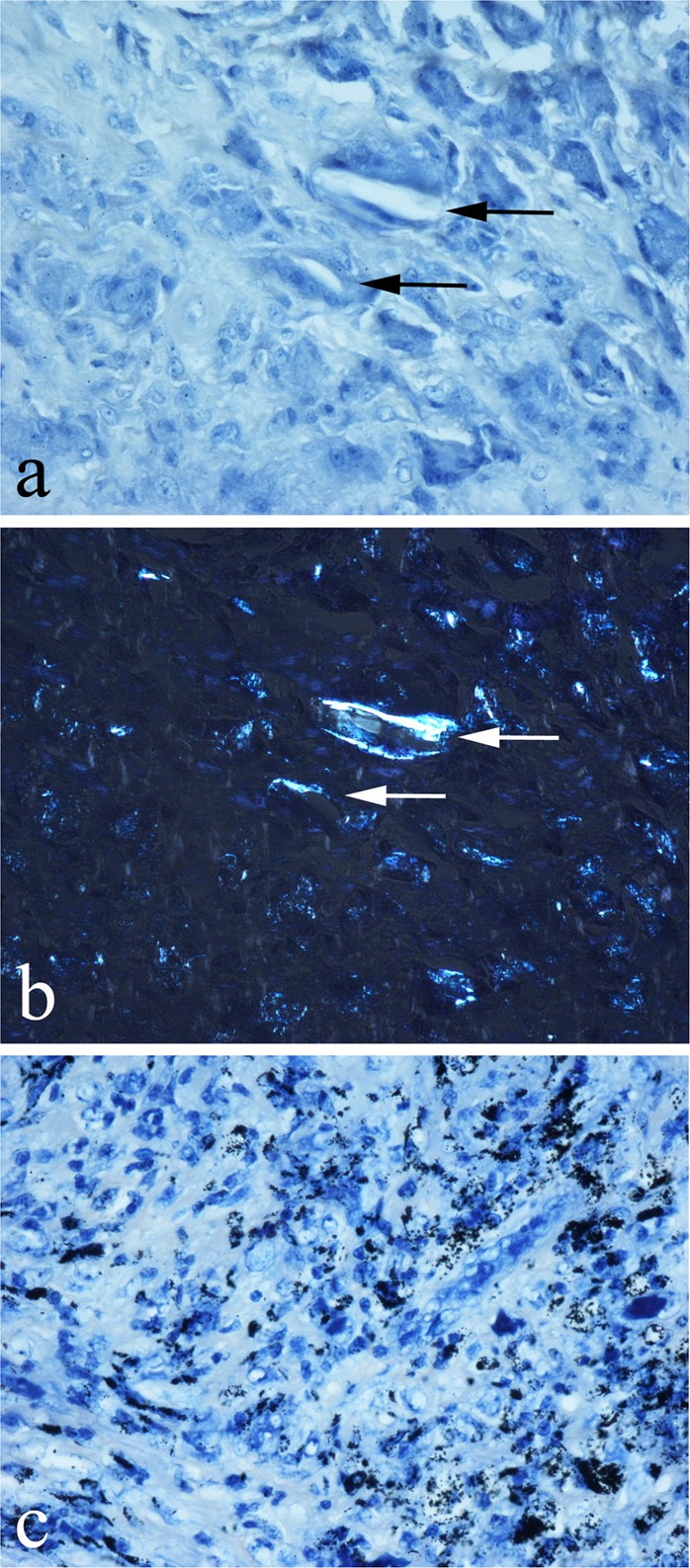
Metal-on-metal (MoM) hip arthroplasties produce abundant implant-derived wear debris composed mainly of cobalt (Co) and chromium (Cr). Cobalt-chromium (Co-Cr) wear particles are difficult to identify histologically and need to be distinguished from other wear particle types and endogenous components (e.g., haemosiderin, fibrin) which may be present in MoM periprosthetic tissues. In this study we sought to determine whether histological stains that have an affinity for metals are useful in identifying Co-Cr wear debris in MoM periprosthetic tissues. Histological sections of periprosthetic tissue from 30 failed MoM hip arthroplasties were stained with haematoxylin-eosin (HE), Solochrome Cyanine (SC), Solochrome Azurine (SA) and Perls' Prussian Blue (PB). Sections of periprosthetic tissue from 10 cases of non-MoM arthroplasties using other implant biomaterials, including titanium, ceramic, polymethylmethacrylate (PMMA) and ultra-high molecular weight polyethylene (UHMWP) were similarly analysed. Sections of 10 cases of haemosiderin-containing knee tenosynovial giant cell tumour (TSGCT) were also stained with HE, SC, SA and PB. In MoM periprosthetic tissues, SC stained metal debris in phagocytic macrophages and in the superficial necrotic zone which exhibited little or no trichrome staining for fibrin. In non-MoM periprosthetic tissues, UHMWP, PMMA, ceramic and titanium particles were not stained by SC. Prussian Blue, but not SC or SA, stained haemosiderin deposits in MoM periprosthetic tissues and TSGT. Our findings show that SC staining (most likely Cr-associated) is useful in distinguishing Co-Cr wear particles from other metal/non-metal wear particles types in histological preparations of periprosthetic tissue and that SC reliably distinguishes haemosiderin from Co-Cr wear debris.
金属对金属(MoM)髋关节置换术后会产生大量的植入物衍生的磨损碎屑,主要由钴(Co)和铬(Cr)组成。钴铬(Co-Cr)磨损颗粒在组织学上难以识别,需要与其他磨损颗粒类型和内源性成分(如血铁黄素、纤维蛋白)区分开来,这些成分可能存在于 MoM 假体周围组织中。在这项研究中,我们试图确定对金属具有亲和力的组织学染色是否有助于识别 MoM 假体周围组织中的 Co-Cr 磨损颗粒。对 30 例失败的 MoM 髋关节置换术后假体周围组织的组织学切片进行苏木精-伊红(HE)、Solochrome Cyanine(SC)、Solochrome Azurine(SA)和 Perls' Prussian Blue(PB)染色。对 10 例使用其他植入物生物材料(包括钛、陶瓷、聚甲基丙烯酸甲酯(PMMA)和超高分子量聚乙烯(UHMWP))的非 MoM 关节置换术的假体周围组织的切片进行了类似的分析。还对 10 例含血铁黄素的膝关节腱鞘巨细胞瘤(TSGCT)切片进行了 HE、SC、SA 和 PB 染色。在 MoM 假体周围组织中,SC 染色吞噬性巨噬细胞和浅层坏死区中的金属碎屑,而这些区域对纤维蛋白的三色染色很少或没有。在非 MoM 假体周围组织中,UHMWP、PMMA、陶瓷和钛颗粒不会被 SC 染色。普鲁士蓝,但不是 SC 或 SA,染色 MoM 假体周围组织和 TSGT 中的血铁黄素沉积物。我们的研究结果表明,SC 染色(可能与 Cr 相关)有助于在假体周围组织的组织学制备中区分 Co-Cr 磨损颗粒与其他金属/非金属磨损颗粒类型,并且 SC 可靠地区分血铁黄素和 Co-Cr 磨损颗粒。