Perino G, Sunitsch S, Huber M, Ramirez D, Gallo J, Vaculova J, Natu S, Kretzer J P, Müller S, Thomas P, Thomsen M, Krukemeyer M G, Resch H, Hügle T, Waldstein W, Böettner F, Gehrke T, Sesselmann S, Rüther W, Xia Z, Purdue E, Krenn V
1Department of Pathology and Laboratory Medicine, Hospital for Special Surgery, 535 E 70th Street, New York, NY 10023 USA.
2Medizinische Universität Graz, Institut für Pathologie, Graz, Austria.
BMC Clin Pathol. 2018 Aug 25;18:7. doi: 10.1186/s12907-018-0074-3. eCollection 2018.
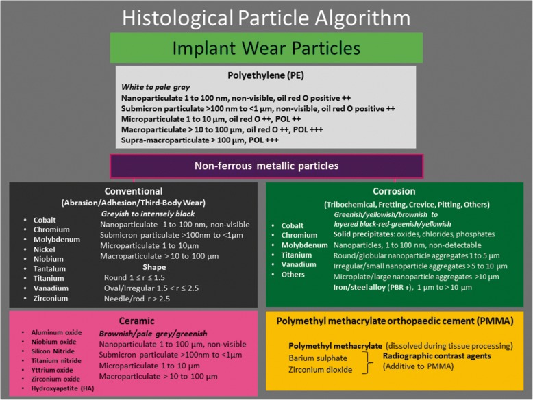
The identification of implant wear particles and non-implant related particles and the characterization of the inflammatory responses in the periprosthetic neo-synovial membrane, bone, and the synovial-like interface membrane (SLIM) play an important role for the evaluation of clinical outcome, correlation with radiological and implant retrieval studies, and understanding of the biological pathways contributing to implant failures in joint arthroplasty. The purpose of this study is to present a comprehensive histological particle algorithm (HPA) as a practical guide to particle identification at routine light microscopy examination.
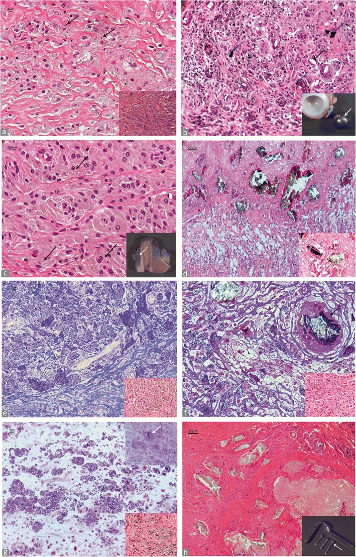
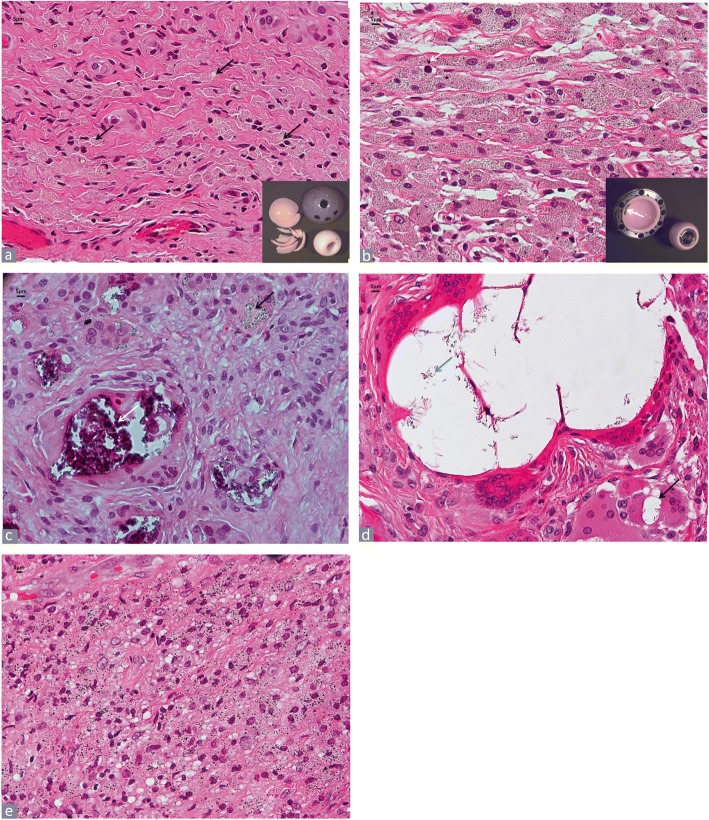
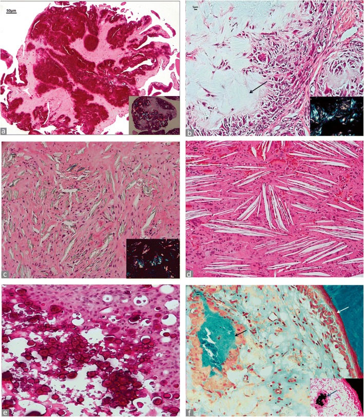
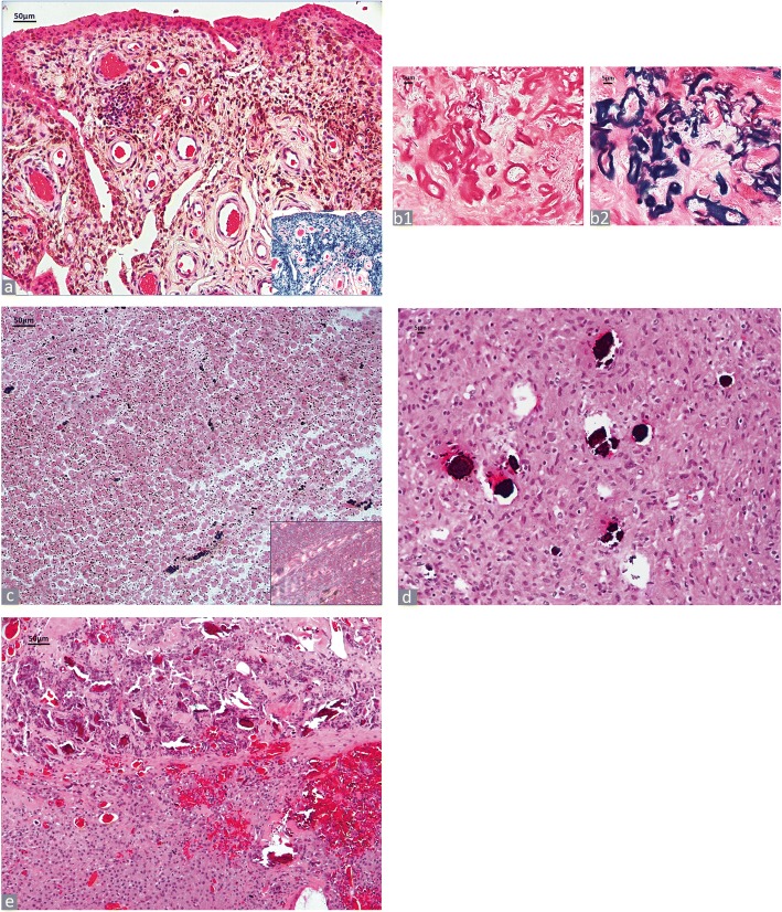
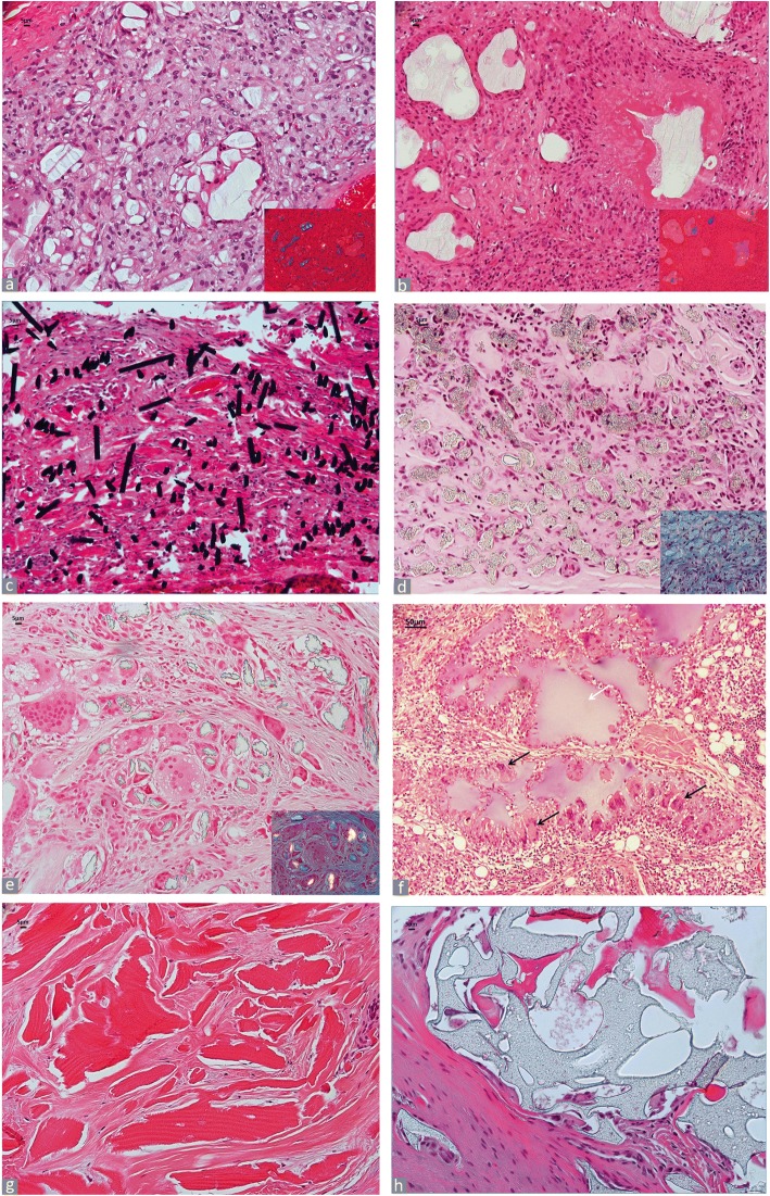
The cases used for particle analysis were selected retrospectively from the archives of two institutions and were representative of the implant wear and non-implant related particle spectrum. All particle categories were described according to their size, shape, colour and properties observed at light microscopy, under polarized light, and after histochemical stains when necessary. A unified range of particle size, defined as a measure of length only, is proposed for the wear particles with five classes for polyethylene (PE) particles and four classes for conventional and corrosion metallic particles and ceramic particles.
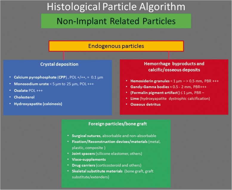
All implant wear and non-implant related particles were described and illustrated in detail by category. A particle scoring system for the periprosthetic tissue/SLIM is proposed as follows: 1) Wear particle identification at light microscopy with a two-step analysis at low (× 25, × 40, and × 100) and high magnification (× 200 and × 400); 2) Identification of the predominant wear particle type with size determination; 3) The presence of non-implant related endogenous and/or foreign particles. A guide for a comprehensive pathology report is also provided with sections for macroscopic and microscopic description, and diagnosis.
The HPA should be considered a standard for the histological analysis of periprosthetic neo-synovial membrane, bone, and SLIM. It provides a basic, standardized tool for the identification of implant wear and non-implant related particles at routine light microscopy examination and aims at reducing intra-observer and inter-observer variability to provide a common platform for multicentric implant retrieval/radiological/histological studies and valuable data for the risk assessment of implant performance for regional and national implant registries and government agencies.
识别植入物磨损颗粒和非植入物相关颗粒,以及表征假体周围新滑膜、骨和滑膜样界面膜(SLIM)中的炎症反应,对于评估临床结果、与放射学和植入物取出研究的相关性以及理解导致关节置换术中植入物失败的生物学途径具有重要作用。本研究的目的是提出一种全面的组织学颗粒算法(HPA),作为常规光学显微镜检查中颗粒识别的实用指南。
用于颗粒分析的病例是从两个机构的档案中回顾性选取的,代表了植入物磨损和非植入物相关颗粒谱。所有颗粒类别均根据其在光学显微镜下、偏振光下以及必要时组织化学染色后观察到的大小、形状、颜色和特性进行描述。对于磨损颗粒,提出了一个统一的粒径范围(仅定义为长度测量),其中聚乙烯(PE)颗粒分为五类,传统金属颗粒、腐蚀金属颗粒和陶瓷颗粒分为四类。
对所有植入物磨损颗粒和非植入物相关颗粒按类别进行了详细描述和说明。提出了一种用于假体周围组织/SLIM的颗粒评分系统如下:1)在光学显微镜下通过低倍(×25、×40和×100)和高倍(×200和×400)两步分析来识别磨损颗粒;2)确定主要磨损颗粒类型并测定其大小;3)是否存在非植入物相关的内源性和/或外源性颗粒。还提供了一份全面病理报告的指南,包括宏观和微观描述以及诊断部分。
HPA应被视为假体周围新滑膜、骨和SLIM组织学分析的标准。它为常规光学显微镜检查中识别植入物磨损颗粒和非植入物相关颗粒提供了一个基本的标准化工具,旨在减少观察者内和观察者间的变异性,为多中心植入物取出/放射学/组织学研究提供一个共同平台,并为区域和国家植入物登记处及政府机构进行植入物性能风险评估提供有价值的数据。